
Abstract
Introduction:
Lumbar hernia defects were first described by Barbette in 1672 and later published by Degarangeot in 1731. 1 The Petit triangle was believed to be the origin of all posterior abdominal wall hernias. In 1866, Grynfeltt, and later Lesshaft in 1870, independently described a superior lumbar triangle. 2 These rare flank hernias appear most commonly on the left in the superior lumbar triangle. They are classified as congenital or acquired with the most common being spontaneous, which make up over half of these hernias. The superior lumbar triangle, or the Grynfeltt–Lesshaft triangle, is bordered superiorly by the 12th rib, medially by the erector spinae and quadratus lumborum and laterally by the internal oblique. The floor of the defect is the transversalis fascia. These hernias are typically repaired through an open approach through a retroperitoneal incision, or laparoscopically through a transabdominal approach. 3 This video depicts a modified laparoscopic extraperitoneal repair using balloon dissection in the posterior intramuscular space.
Materials and Methods:
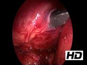
A 76-year-old male presented with a 2-month history of pressure and pain while straining. These symptoms were exacerbated by flexion, twisting, and straining while using the restroom. CT imaging depicted a right-sided superior lumbar triangle hernia with perinephric fat protruding through the defect. Operatively, the patient was positioned in the left lateral decubitus position. The hernia defect, 12th rib, 11th rib, and the subcostal region were marked on the patient. A 12 mm incision was made in the midaxillary line between the iliac crest and the 12th rib. The subsequent dissection split the external and internal obliques bluntly. A plane was developed beneath the internal oblique and above the transversalis fascia staying superficial to the peritoneum and abdomen. A balloon dissector was inserted into the space and inflated under direct observation. Using blunt dissection, the hernia contents were reduced and the facial defect was delineated. The facial edges were closed with interrupted 0 silk and reinforced with polypropylene mesh.
Results:
The operation took 97 minutes with minimal blood loss. The patient was observed overnight and discharged home on postoperative day 1, tolerating a regular diet. He underwent repeat imaging 8 months postoperatively that showed no recurrence of the right-sided flank hernia.
Conclusion:
Lumbar hernias are rare with no standard approach to their repair. This technique allows for a minimally invasive repair while avoiding the risk of intra-abdominal injuries associated with a transabdominal approach.
No competing financial interests exist
.
Runtime of video: 7 mins 40 secs
This video was first presented as a podium presentation at the American College of Surgeons 2019 Clinical Congress.


Keywords
Get full access to this article
View all access options for this article.