
Abstract
Introduction:
Morgagni hernias account for 1.5%–6% of all congenital diaphragmatic defects. 1 The majority of Morgagni hernias are unilateral, usually right sided with 2% being bilateral. 2 Most patients are asymptomatic with a third presenting with respiratory or gastrointestinal symptoms, and there is a strong association with Downs syndrome (26%) and congenital heart defects (34.8%). Most are amenable to primary laparoscopic repair but bilateral Morgagni hernias (BMH) is more challenging with case reports describing the need for a mesh repair of the defect. 1 –3 Demonstrated here are the technical steps in a primary repair of a BMH without a mesh.
Material and methods:
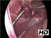
A 2-year-old Downs syndrome girl under cardiology care was referred with a diaphragmatic hernia. X-rays revealed a large central Morgagni hernia and cardiac echo confirmed fitness for surgery. The patient was placed in French position and an umbilical camera and two paraumbilical 5 mm ports were utilized. A large BMH containing transverse colon and small bowel was noted and contents were reduced. Repair proceeded with percutaneous placement of Prolene 2/0 sutures through the anterior abdominal wall through two 1 cm incisions on either side of the midline inferior to the xiphoid process. These were retrieved intra-abdominally and passed through the posterior fold of the diaphragm before being passed out through the same incision and held with forceps. A series of three sutures were placed on each side, and once in position, the anticipated peritoneal contact margins of the diaphragm and anterior abdomen were roughened with monopolar diathermy to facilitate adhesion. The hernia sac was left
Results:
Surgery was accomplished with minor distortion of the anterior chest and abdominal wall. The patient was fed on recovery and a postoperative chest X-ray showed a good result. Discharge was on day 4. At 6 months, the patient remains well with no recurrence of the BMH.
Conclusion:
Critical steps in BMH repair are an incremental closure starting laterally and finishing medially where tension is the greatest, freshening of margins of the defect to consolidate the repair, preferential extracorporeal knotting with supplementary intracorporeal suturing to manage residual small defects, and the use of exclusively nonabsorbable sutures to ensure durability of the repair. Because of strict adhesion to the pericardium, the hernia sac is best left
Runtime of video: 5 mins 46 secs


Get full access to this article
View all access options for this article.