
Abstract
Introduction:
Although 3 and 5 mm port-site hernias are rare in minimally invasive surgery, the incidence is higher in young children and infants possibly because of a thinner abdominal wall and smaller thinner omentum. 1 –4 In our experience, hernias of 3 and 5 mm trocar sites frequently occur in the early postoperative period and require additional operative intervention to reduce the herniated abdominal contents and repair the fascial defect. To prevent the occurrence of 3 and 5 mm port-site hernias and the need for additional procedures in young infants, we have developed a novel technique of port-site closure utilizing laparoscopic assistance.
Materials and Methods:
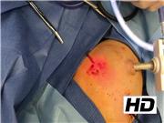
The video shows the incidence of port-site hernias and describes hernia-associated adverse outcomes in the pediatric population. The surgeon demonstrates the port-site closure technique using an RB-1 needle; the video displays both internal (laparoscopic) and external images, as well as an animation of the technique.
Results and Conclusions:
At our institution, we have had no port-site hernias of 3 or 5 mm trocar sites since employing this technique. Laparoscopic assistance enabled better observation of the facial edges and improved precision of the suture placement in the subcutaneous tissue. We recommend routine use of this technique for 3 and 5 mm port-site closures in infants <6 months of age.
No competing financial interests exist.
Runtime of video: 3 mins 48 secs


Get full access to this article
View all access options for this article.