
Abstract
Introduction:
Diaphragm eventration from a phrenic nerve injury after congenital heart surgery is a well-recognized complication. Treatment of the eventration is diaphragm plication if there has been no recovery of the diaphragm function. 1 This has been performed thoracoscopically in children, typically with a single row of nonabsorbable suture without pledgets. 2 Some pediatric surgeons have expressed reluctance in performing plications thoracoscopically, for concern that the single row of sutures without pledgets is not as durable and can pull suture through the muscle. I present a technique of a thoracoscopic diaphragm plication using horizontal mattress suture with pledgets. 3
Materials and Methods:
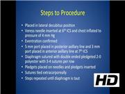
A 9-month-old boy with hypoplastic heart syndrome underwent a Glenn procedure. Postoperatively, he had difficulty weaning oxygen and was tachypneic. A chest X-ray showed an elevated right hemidiaphragm. An ultrasonography and fluoroscopy showed paradoxical motion of the right diaphragm, confirming an eventration. This did not improve after 21 days and a thoracoscopic diaphragm plication was undertaken. The patient was placed in the lateral decubitus position with the right side up. Three ports were used. A 4 mm port was placed in the mid axillary line at the sixth intercostal space and a 4 mm 30° thoracoscope was used. A 3 mm port is placed in the anterior axillary line and a 5 mm port is placed at the posterior axillary line at the seventh intercostal space.
After confirming the diaphragm eventration, 2-0 polyester suture is inserted through the 5 mm port and a horizontal mattress suture is performed. The second row of the double-ended suture is placed in the same position a few millimeters lateral to the first row. The pledget that comes preloaded on the suture is pulled into the chest. The needle from the second row is pulled out and placed onto a second pledget. Then the first row needle is pulled out and placed on the same pledget and the pledget is inserted into the chest. A knot pusher is used to tie the two rows together. This allows pledgets on both sides of the rows of sutures. This process is repeated going laterally for 3–4 rows of sutures, bringing the diaphragm to an appropriate tension.
Results:
The postoperative CXR showed the right diaphragm in a good position. The child was extubated on postoperative day 1 and weaned to room air by postoperative day 3.
Conclusions:
A horizontal mattress pledgeted plication of the diaphragm can be performed thoracoscopically. I believe this technique distributes the tension better on the diaphragm muscle. Although pledgets have not routinely been used in other descriptions of thoracoscopic repairs, it does not add much time, is easy to insert, and adds durability to the repair to prevent pulling through of sutures.
No competing financial interests exist.
Runtime of video: 5 mins


Get full access to this article
View all access options for this article.