
Abstract
Background:
Solid pseudopapillary neoplasms of the pancreas are rare in pediatric patients. 1,2 Recently, there have been reports of laparoscopic approaches to the treatment of pseudopapillary neoplasms of the pancreas, including tumor enucleation, distal pancreatectomy, and pancreatoduodenectomy. 3 –5 We herein report a case in which laparoscopic distal pancreatectomy with spleen and vessel preservation was effectively performed to treat a pediatric patient with a huge solid pseudopapillary neoplasm.
Patients and Methods:
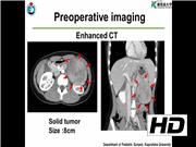
The patient was an 11-year-old girl in whom proteinuria was detected in a routine checkup at her school. A left upper abdominal tumor was incidentally detected by ultrasonography and enhanced computed tomography. The 8 cm tumor was solid and had originated from the pancreas. A solid pseudopapillary neoplasm was suspected based on the imaging findings.
The Operative Findings and Procedure:
Under general anesthesia, the patient was placed in a right semilateral position, and a 10-mm 30° laparoscope was inserted through an umbilical incision using the open 12-mm Hasson trocar method. The huge tumor was easily recognized in the left upper abdomen. Three additional 5-mm trocars were inserted in the right upper abdomen (operator's left hand), left side of the umbilicus (operator's right hand), and the left lateral abdomen (assistant). The bursa omentalis was opened using the vessel sealing system, and the tumor was confirmed to have originated from the pancreatic tail. The tumor was located 3 cm laterally from the superior mesenteric vein. Thus, transection of pancreatic body was feasible for tumor resection. To expose the whole tumor, the gastrosplenic ligament was divided using the vessel sealing system. The retroperitoneum was incised at the inferior border of the pancreatic body. The posterior surface of the pancreatic body was carefully dissected with coagulating small branch vessels. The drainage vein of the tumor, which was connected to the splenic tail vein, was ligated using a clip, and divided. The splenic vein was detected and confirmed after tunneling dissection of the pancreatic body. Tiny veins from the splenic vein were coagulated and divided using the vessel sealing system. The retroperitoneum was incised between the superior border of the pancreatic body and splenic vein. The taping of the pancreatic body was performed. The pancreatic body was carefully stapled using a linear stapler and transected without injuring the splenic vessels. The layer of NEOVEIL provides support to the tissue that is being stapled or resected, particularly benefiting fragile tissue such as in the lungs that may otherwise sustain damage during the procedure. The stump of pancreatic body was confirmed to be stapled. The transected distal pancreas was dissected along the splenic vein. The feeding vessel from the splenic artery was also coagulated and divided using the vessel sealing system. The tumor was extracted through a 6-cm Pfannenstiel incision.
Results and Conclusion:
There were no intraoperative or postoperative complications. Pancreatic fistula was not observed. The postoperative course was uneventful. In the present case, laparoscopic distal pancreatectomy with spleen and vessel preservation was a safe and feasible approach to the treatment of a pediatric patient with a huge solid pseudopapillary neoplasm.
Runtime of video: 5 mins 23 secs


Get full access to this article
View all access options for this article.