
Abstract
Introduction:
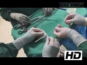
This video describes magnetic resonance imaging-operating room (MRI-OR) guided laparoscopic anorectoplasty utilizing a novel patient positioning apparatus in a 1-year-old male patient. Traditional approaches to imperforate anus include the posterior sagittal anorectoplasty (PSARP) with the recent adopting the laparoscopic assisted approaches. 1 The PSARP involves a midline incision bisecting the sphincter complex, which can lead to scarring and weakness. The laparoscopic assisted approach popularized by Georgeson uses a straight trocar to go from the skin to pelvic floor, which may also miss the sphincter complex. 2
Materials and Methods:
Here, we present a technique adopted from Raschbaum et al., with utilization of an MRI-OR for image-guided trocar placement through the sphincter complex. 3,4 We utilized a novel patient apparatus that is both MRI compatible and allows for proper patient positioning for trajectory of the needle. During the procedure, a percutaneous needle is placed through the sphincter complex under direct observation, and serial MRI-guided adjustments are made to ensure proper location. Once this is completed, the patient receives a laparoscopic mobilization and dissection of the distal rectum, ligation of fistula tract, and placement of anorectral trocar through the previous tract. The neorectum is then brought through the tract and sutured to the skin.
Conclusions:
This highlights the ability to utilize image guidance for preservation of the sphincter complex during a laparoscopic anorectoplasty for imperforate anus.
No competing financial interests exist.
Runtime of video: 5 mins 2 secs
Previously presented at the International Pediatric Endosurgery Group (IPEG) annual meeting, in London, UK, July 2017.


Get full access to this article
View all access options for this article.