
Abstract
Introduction:
Symptomatic malrotation occurs in ∼1/6000 live births. 1 The narrow root of the mesentery makes the bowel prone to volvulus, which can lead to small bowel ischemia. This problem can be managed surgically through a laparoscopic Ladd procedure.
Materials and Methods:
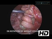
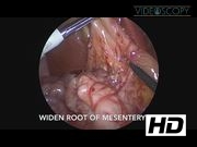
This video shows a stepwise laparoscopic Ladd procedure done in a 13-day old female infant who presented with worsening bilious emesis and ultrasound findings suspicious of malrotation and midgut volvulus. Access to the right lower quadrant was achieved by a modified Hasson technique with a Step™ trocar (Auto-Suture) placed through a 5 mm incision in the umbilicus. Pneumoperitoneum was established using 5 L/min of carbon dioxide under 10 mm Hg of pressure. A 30° telescope camera was inserted into the peritoneal cavity for proper observation. Next, two incisions were made in the lower abdomen. A 3 mm port was placed in the lower right abdomen and a 5 mm port was placed in the lower left abdomen with Step trocars (Auto-Suture). First, important structures were identified. The duodenum and cecum were located in the right upper quadrant with Ladd bands stretching across both structures and attaching to the right lateral abdominal wall. The midgut volvulus was then identified and approximated to be around 1.5 turns. Using the bowel grasper, the volvulus was derotated in an anticlockwise direction and restored to its normal anatomical position. All of the small bowel was examined and determined to be completely viable. Next, hook diathermy was used to release the Ladd bands, freeing the cecum and right side of the colon from the lateral abdominal wall. The root of the mesentery was then widened by dividing the Ladd bands between the cecum and duodenum. All of the small bowel was then placed on the right side of the abdomen, whereas all of the colon was placed on the left side of the abdomen. Once the peritoneal cavity had been examined, the appendix was then located and its mesentery was taken down. The appendix was ligated with two 3′0 Vicryl ties, resected, and removed through a port site. Finally, the stomach was insufflated to rule out an intrinsic duodenal obstruction. Air passed easily into the stomach and across the duodenum, confirming patency. Ports were withdrawn after desufflation of the peritoneal cavity. The umbilical fascia was closed with 2-0 Vicryl UR-6 single suture. All other incision sites were closed with Dermabond glue.
Results and Conclusions:
There were no postoperative complications. The patient began tolerating feeds on POD 5 and was discharged on POD 7. At her 4-week and 6-month follow-up visits, there was no recurrence of midgut volvulus.
No competing financial interests exist.
Runtime of video: 7 mins 46 secs

Get full access to this article
View all access options for this article.