
Abstract
Introduction:
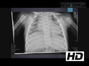
Isomerism is a rare congenital condition in which one side of the body is a mirror image of the opposite side of the body. Abdominal manifestations include abnormal stomach positioning with possible malrotation of the intestines. 1 If malrotation occurs, a laparoscopic Ladd procedure is performed.
Materials and Methods:
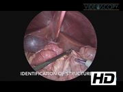
This video shows a laparoscopic Ladd procedure done in a 9-month-old male with right-sided isomerism and asymptomatic intestinal malrotation. Access to the right lower quadrant was achieved by a modified Hasson technique with a Step™ trocar (Auto-Suture) placed through a 5 mm incision in the umbilicus. Pneumoperitoneum was established using 5 L/minute of carbon dioxide under 12 mm Hg of pressure. A 30° telescope camera was inserted into the peritoneal cavity for proper observation. Next, two 3 mm incisions were made in the lower right and left abdominal quadrants. A 3 mm port was placed on the right and a 5 mm port was placed on the left with Step trocars (Auto-Suture). Important structures were immediately identified. The liver was located in the center of the abdomen and the spleen was absent, suggestive of right-sided isomerism. The attachment between the splenic flexure of the colon and the left lateral abdominal wall was divided. The colon was then reflected downwards, exposing the centrally located stomach. Duodenal loops were released from the Ladd bands, allowing for observation of the pancreas lying inside of the C-loop of the duodenum. While looking for the distal portion of the jejunum, it was discovered that the entire jejunum was trapped retroperitoneally, creating a mesocolic hernia. Using hook diathermy, the entire bowel was released down to the ileocecal region, which was in its normal anatomical location in the right lower quadrant. The appendix appeared normal. Using hook diathermy, the mesentery of the appendix was taken down. The appendix was ligated with two 3′0 Vicryl ties, resected, and removed through a port site. All of the small bowel was placed on the right side of the abdomen, and all of the large bowel was placed on the left. Finally, the stomach was insufflated to rule out an intrinsic duodenal obstruction. Air passed easily into the stomach and across the duodenum, confirming patency. The bowel appeared completely viable. Ports were withdrawn after desufflating the pneumoperitoneum. The umbilical port site was closed with 2-0 Vicryl UR-6 single suture. All other port sites were closed with Dermabond glue.
Results and Conclusions:
There were no postoperative complications. The patient began tolerating feeds on post-operative day #3 and was discharged on post-operative day #4.
No competing financial interests exist.
Runtime of video: 5 mins 51 secs

Get full access to this article
View all access options for this article.