
Abstract
Introduction:
Laparoscopic intragastric surgery was demonstrated to remove the lesion of posterior wall of stomach. 1 We performed this procedure for the pediatric rare disease not only to ensure the minimally invasiveness and good cosmetic appearance but also to preserve the cardia function of stomach. In the video, we present our intragastric resection for gastric tumor under augmented reality navigation system.
Materials and Methods:
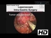
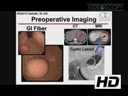
A 12-year-old boy was admitted due to hematemesis. Gastrointestinal (GI) fiberscope revealed the 4 cm size submucosal tumor located at the posterior wall. Located position was near the EC junction. The tumor was cystic type and seemed to be benign lesion. Hence, we planned to perform the intragastric surgery to preserve the cardia function of stomach. We also set up this procedure under augmented reality navigation system to prevent the around organ injury. 2 Initially, GI fiberscope was introduced orally to determine the tumor. Next, laparoscope was introduced through the umbilical incision using the open Hasson's method and pneumoperitoneum was established. And then, we used the Funada-kit II (Create Medic Co., Kanagawa, Japan) device with two parallel needles to puncture the stomach and assist suturing the anterior gastric wall to the anterior abdominal wall. In this procedure, all three trocars are placed in the gastric lumen, penetrating both the abdominal and stomach walls to perform a laparoscopic removal of gastric lesions under the observation of GI fiberscope. The operation is then carried out in the gastric lumen with currently available laparoscopic instruments and laparoscopic monitoring. 3 Laparoscopic intragastric surgery requires a pneumostomach, which is created by CO2 insufflations through the GI fiberscope. 4 Volume images were reconstructed by three-dimensional (3D) viewer application. We used an optical tracking system for registration between volume image and body surface markers. The augmented reality visualization was superimposed preoperative 3D computed tomography (CT) images onto captured laparoscopic live images. Preoperative 3D CT was performed under condition of maximum swallowing air. This is because of the mimicking the situation of pneumostomach during intragastric surgery. Retroperitoneal organs, such as splenic artery and vein, spleen, and pancreas, did not show much deformation and deviation from the data of our previous clinical experiments. 2 The operator recognized the hidden vascular variation of the splenic artery and vein, spleen, and pancreas by overlaying an image onto a laparoscopic live image. The tumor was resected by ultrasonically activated device using intragastric surgery. Esophagocardial (EC) junction was kept without injury. Defect of gastric wall was closed under laparoscopic view.
Results:
There were no intra- and postoperative complications. Pathological diagnosis revealed that the tumor was bronchogenic cyst. The patient had good clinical course after operation.
Conclusion:
Laparoscopic intragastric surgery is feasible and safe and effective for the pediatric patients. This approach is useful for the tumor lesion of posterior wall and near EC junction to preserve the organ function and to prevent the injury of EC junction. No relevant articles were found in the literature search, and this is the first report referring to the application of intragastric surgery for child case.
Runtime of video: 3 mins 52 secs

Get full access to this article
View all access options for this article.
References
Supplementary Material
Please find the following supplemental material available below.
For Open Access articles published under a Creative Commons License, all supplemental material carries the same license as the article it is associated with.
For non-Open Access articles published, all supplemental material carries a non-exclusive license, and permission requests for re-use of supplemental material or any part of supplemental material shall be sent directly to the copyright owner as specified in the copyright notice associated with the article.