
Abstract
Introduction:
Rectal atresia is rare, comprising only 1% of all anorectal malformations. Initial evaluation reveals a normal appearing anus located within the sphincter mechanism. A blind ending pouch is 3 cm from the anal verge. The anal canal and internal and external sphincters are well developed and there is no associated fistula. No technique has been frequently reproduced for this rare anomaly, and only one laparoscopic report exists. We present a laparoscopic and transanal approach for rectal atresia repair that provides minimal disturbance of the anal anatomy and achieves postoperative continence.
Materials and Methods:
A term, 3.5 kg male presented with failure to pass meconium and progressive abdominal distension. On physical examination, the external rectum appeared normal; however, rectal examination revealed a palpable, blind ending, web at 3 cm from the anal verge. The physical examination was otherwise normal. A contrast enema revealed a complete transverse web across the rectum. Spinal and abdominal ultrasonography and magnetic resonance imaging of the abdomen and pelvis confirmed the absence of a presacral mass or renal anomaly. The patient underwent colostomy and mucous fistula to relieve the initial obstruction. A mucous fistulogram confirmed the anatomy of the web and the absence of a fistula.
Operation:
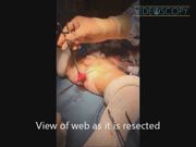
At 6 weeks, the infant underwent laparoscopic and transanal repair of rectal atresia. Under general anesthesia, the patient was prepared in the Trendelenburg position. A foley catheter was placed. The Pena stimulator confirmed the rectum was within the sphincter complex. Three 5 mm laparoscopic trocars were inserted at the umbilicus and right and left upper quadrants, respectively. Carbon dioxide insufflation was maintained at 8 mm Hg. With the 5 mm laparoscopic camera, the indentation of the web was confirmed after placing a Hegar dilator into the rectum. Circumferential dissection proceeded around the rectal attachments. Transanal resection of the web proceeded with exposure from the Lonestar retractor. A submucosal dissection commenced at 1 cm above the dentate line. The sphincters were preserved as the web was removed in the submucosal plane. The normal rectum from above the web was sutured in place at 1 cm above the dentate line with interrupted Vicryl 4/0 sutures. The anastamosis had no tension, well vascularized, and correctly oriented. Operative time was 85 minutes. The patient was discharged 48 hours after surgery. There were no complications. A dilation program was completed between 2 weeks and 3 months after the operation. At 6 weeks the colostomy was closed. At 18 months, the patient has excellent bowel function.
Conclusion:
The laparoscopic and transanal approach for rectal atresia is a unique procedure with several advantages. All elements that contribute to continence, including the rectal mucosa and the internal and external sphincters, are preserved. The laparoscopic approach causes less abdominal trauma than previously described abdominal approaches. The transanal dissection causes less sphincter disruption than the posterior-sagittal approach. Complications such as bleeding, twisting of the pull through, and inadvertent damage to pelvic structures, as reported with straight pull through, are avoided. We report a satisfactory outcome in one patient.
Runtime of video: 5 mins


Get full access to this article
View all access options for this article.
Supplementary Material
Please find the following supplemental material available below.
For Open Access articles published under a Creative Commons License, all supplemental material carries the same license as the article it is associated with.
For non-Open Access articles published, all supplemental material carries a non-exclusive license, and permission requests for re-use of supplemental material or any part of supplemental material shall be sent directly to the copyright owner as specified in the copyright notice associated with the article.