
Abstract
Introduction:
Leiomyoma is a benign bladder tumor accounting for ∼0.43% of all bladder tumors. 1,2 There are ∼200 reported cases, with ∼75% occurring in females during their third to sixth decades of life 3 , making this tumor extremely rare in the pediatric population. This video demonstrates an enucleation of bladder wall leiomyoma using single-incision pediatric endoscopic surgery (SIPES) with a glove access port in a pediatric patient.
Materials and Methods:
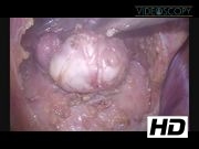
The patient is a 16-year-old female who initially presented to the emergency department with a 24-hour history of abdominal pain and fevers. On examination, she had lower midline abdominal wall tenderness without guarding. Her white blood cell was 6.0 k/mm3 and the abdominal ultrasound was unable to visualize the appendix; therefore, she was admitted for observation to rule out early appendicitis. Given her persistent symptoms, she underwent an abdominal computed tomography (CT) scan with contrast. The CT scan revealed a 3 × 2 × 3 cm lobulated soft tissue mass intimately related to the anterior wall of the urinary bladder. Radiologic findings were believed to be consistent with an urachal remnant; however, a primary tumor could not be ruled out. Thus, an abdominal ultrasound was obtained to better define the mass, which confirmed a homogenous soft tissue mass consistent with an urachal cyst. Accordingly, the patient was treated with a 7-day course of Levaquin for a presumed infected urachal remnant. Following treatment, surgical resection was planned to prevent future episodes of infection. A SIPES approach was used. A single-incision umbilical port was constructed using an extra small AlexisTM (Applied Medical, Rancho Santa Margarita, CA) wound protector and a sterile 6.5 orthopedic glove. The thumb was used for insufflation, and the fingers for the laparoscope and 5-mm instruments. On inspection, a mass was protruding from the dome of the bladder. However, the mass did not have a typical appearance for an urachal remnant, as there was no tract extending to the umbilicus. Enucleation of the mass was carried out using monopolar electrocautery and blunt dissection without entering the bladder. After complete resection, the bladder was reconstructed in a single layer using a 2-0 V-LocTM suture (Covidien, Minneapolis, MN). The bladder was then filled with saline and no extravasation was identified. The peritoneum over the bladder was reapproximated using a single 2-0-Vicryl (Ethicon, Somerville, NJ). The fascia was closed using a 0-PDS and the skin was closed with a 4-0-Monocryl (Ethicon).
Results:
The total procedure time was 135 minutes. The patient tolerated the procedure without any complications and was discharged home on postoperative day 1. Pathology revealed a spindle cell tumor with clear margins, consistent with a benign leiomyoma. It is reported that ∼13% of patients present with pain. 3 However, there is no correlation between fevers and leiomyomas. After discharge, her only complaint was transient mild bladder spasms, which were treated successfully with a course of oxybutynin. The abdominal pain and fevers experienced preoperatively resolved.
Conclusion:
In conclusion, SIPES is an effective and safe approach for the resection of bladder wall leiomyomas in children.
This video will be presented at IPEG's 23rd Annual Congress for Endosurgery in Children.
No competing financial interests exist.
Runtime of video: 4 mins 17 secs


Keywords
Get full access to this article
View all access options for this article.
References
Supplementary Material
Please find the following supplemental material available below.
For Open Access articles published under a Creative Commons License, all supplemental material carries the same license as the article it is associated with.
For non-Open Access articles published, all supplemental material carries a non-exclusive license, and permission requests for re-use of supplemental material or any part of supplemental material shall be sent directly to the copyright owner as specified in the copyright notice associated with the article.