
Abstract
Background:
We present a rare case of a metanephric adenoma, excised using an ultrasound (US) guided, nephron-sparing, laparoscopic approach.
Introduction:
A metanephric adenoma is a rare, benign tumor of the kidney, with only 80 cases reported in the surgical literature. 1 Surgical excision is curative and does not require removal of the entire kidney. 2 –4 However, it can be difficult to localize the tumor laparoscopically. We present an US guided, transperitoneal, nephron-sparing approach to this lesion.
Case History:
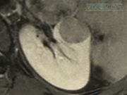
An otherwise fit and well 16-year-old female presented to the emergency department with abdominal pain. The only medical history of note was a normal renal ultrasound performed at 18 months of age, following a urinary tract infection. An abdominal ultrasound revealed a 2.4-cm isoechoic mass in the interpolar region of the left kidney. A subsequent MRI confirmed a solid renal lesion with no associated lymphadenopathy. Urinary cytology was normal. Following discussion at the local tumor advisory meeting, it was decided to undertake nephron-sparing surgery to obtain a histological diagnosis, as renal cell carcinoma could not be excluded.
Procedure:
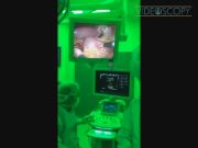
Using a transperitoneal approach, the kidney was exposed. Intraoperative ultrasound was used to delineate the lesion. The arterial and vascular supply to the tumor were then clipped and divided. Using the Enseal, the devascularized portion of the kidney containing the tumor was then removed. The cut surface was then oversown and sprayed with tissue sealant. The patient made an uneventful recovery and was discharged 2 days following the procedure. An ultrasound performed 1 and 6 months postoperatively, has shown no urinoma or tumor recurrence. Renal function 6 months postoperatively was normal at 59 μM compared to 52 preoperatively (normal = 50–100 μM).
Results:
The tumor was successfully removed, with clear excision margins, without the need for total nephrectomy.
Discussion:
The use of intraoperative ultrasound allowed for accurate and confident identification of the small target lesion. By removing the minimum amount of renal tissue, only a small number of vessels required division. This avoided the need for renal vessel clamping and the associated risks of warm ischemia time. 5,6 The use of the Enseal bipolar sealing device allowed for rapid excision of the remaining tissue and excellent hemostasis. Combined with the US guidance, this allowed for the preservation of the adjacent renal collecting system, avoiding the need for total nephrectomy. Urinary leak from the cut kidney surface was effectively prevented by oversowing the area and using tissue sealant.
Iain Hennessey: Covidien Minimal Access Scholarship.
Runtime of video: 5 mins 58 secs

Keywords
Get full access to this article
View all access options for this article.
References
Supplementary Material
Please find the following supplemental material available below.
For Open Access articles published under a Creative Commons License, all supplemental material carries the same license as the article it is associated with.
For non-Open Access articles published, all supplemental material carries a non-exclusive license, and permission requests for re-use of supplemental material or any part of supplemental material shall be sent directly to the copyright owner as specified in the copyright notice associated with the article.