
Abstract
Introduction:
Robotic approach has been increasingly applied to the surgical resection of distal rectal cancer. All reports from the advocators on the use of robotic assistance in rectal surgeries have confirmed its safety, efficacy, and good functional outcomes. 1 –5 Currently, however, there is no denying that robotic technology in rectal cancer surgery is still in its infancy, which is reflected in the limited number of publications on this topic, and the variability of robotic techniques ranging from the use of the robot in a single cart position (single docking) or two different cart positions (dual docking), to the use of hybrid procedure. In the present study, we test the technical feasibility of our individual robotic approach, the three-armed robotic abdominoperineal resection (APR), for the treatment of distal rectal cancer. We hypothesize that by using three Da Vinci® robotic arms (Intuitive Surgical, Sunnyvale, CA), the APR procedure can be performed as efficiently and efficaciously as by using the standard four robotic arms.
Methods:
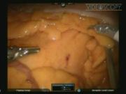
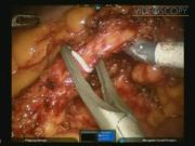
Patients with distal rectal cancer, defined as the tumor location being within 5 cm or below the first Houston valve above anal verge, were recruited onto this study and subjected to the present three-armed robotic APR procedure. The baseline clinicopathologic data and the surgical outcomes of patients were prospectively followed up and evaluated. Details of the surgical technique are presented in the attached streaming video. Briefly, five abdominal ports were set: 12-mm camera port placed 3 cm to the right and 3 cm above umbilicus; 12-mm port right lower quadrant (mid-clavicular line) through which is telescoped an 8-mm robotic port designated as R1 for the right robotic working arm (The 8-mm port can be removed to place an endostapler); 8-mm port left upper quadrant just to the right of the mid-clavicular line midway between umbilicus and left subcostal region designated as R2 for the left robotic working arm; 12-mm assistant port right lateral mid-abdomen for retracting and suctioning by an assistant; 8-mm port left lower quadrant placed at the same height and positioned as the right lower quadrant port for counter-traction by another assistant. The robotic cart was placed in left hip position. During the entire APR procedure, each robotic working arm and assistant instrument was inserted through its respective uniform abdomen port cited above, and the position of the robotic cart was remained unchanged. The robotic dissection sequence was similar to that of APR we performed by traditional laparoscopic surgery 6 , comprising (1) medial-to-lateral mesenteric dissection along the pre-aortic plane; (2) optional ligation and transection of inferior mesenteric vein; (3) N3 lymph node dissection over the root of inferior mesenteric artery; (4) low ligation of vascular pedicle with preservation of left colic artery and autonomic nerve plexus; (5) mobilization of sigmoid and descending colon; (6) dissection of pre-sacral fascia downward to the anococcygeal rhaphae with preservation of paired hypogastric nerves and pelvic autonomic nerve plexi; (7) incision of the peritoneal reflection laterally and then anteriorly; and (8) separation of the rectum and the vagina/prostate circumferentially to the level of levators with the appreciation of Denonvilliers' fascia. After completion of the robotic total mesorectal excision (TME), the robotic cart is removed and a colostoma was shaped over the left low abdominal quadrant. Then, we undertook the perineal dissection and finally the rectal cancer was extirpated in cylindrical style.
Results:
A total of 12 patients was accrued within 9-month period commencing February 2012, under the ethical approval of clinical trial in National Taiwan University Hospital. The distribution of age was 59.3 ± 13.1 years (mean ± standard deviation). There were four females and eight males. The body-mass index (BMI) was 28.2 ± 3.0 kg/m2. The physical status (classification of American Society of Anesthesiology) was class I in nine, class II in two, and class III in one patients. The histopathology included adenocarcinoma in 11 patients and neuroendocrine tumor in 1. There were 10 patients undergoing preoperative chemoradiation therapy (CCRT) and 2 received surgical intervention directly. The pathologic TNM stage was four patients with complete response after CCRT (stage 0), two patients in stage I, two in stage II, and four in stage III. The operation time was 285.0 ± 99 minutes (including docking time). The median blood loss was 100 mL (range 50–450 mL). There was no major complication. However, perineal wound infection of port sites was detected in two patients. The patients have quick convalescence, as evaluated by the length of postoperative ileus (48.0 ± 12.0 hours), hospitalization (10 ± 2.0 days), and degree of postoperative pain (2.5 ± 0.5 visual analogue scale). The median time for the removal of Foley catheter was 7 days (range 5–21 days). The median time of return to partial activity, full activity, and work was 2.0, 4.0, and 6.0 weeks, respectively. The number of cleared lymph nodes was 16.1 ± 7.2. Besides the expenses covered by the National Bureau of Health Insurance of Taiwan, the consumable payment by patients undergoing a robotic APR was NT$180,000.0 ± 3000.0 (1 U.S. dollars = 29NT$).
Discussion:
The present case series have shown that by using three Da Vinci robotic arms in our clinical setting, the APR procedures can be performed for patients with good technical efficiency, quick functional recovery, and mild disability. Since a complete mobilization of the left colon up to the colonic splenic flexure will not be necessary in most APR cases except when patients with extreme habitus (very tall or very high BMI), the entire extent of dissection can be achieved with three robotic arms and without any change of position of the robotic cart. In our method, the use of three robotic arms can translate into the saving of one-fourth expenses of robotic consumables, compared with the standard four-arm robotic approach; and single docking of the robotic cart can translate into more simple and timesaving for the whole surgical procedure, as compared with multiple docking method. With the above-mentioned dual technical benefits in mind, we therefore suggest that the present three-armed single-docking method should be recommended to patients with rectal cancer requiring an APR, if their body habitus were within normal limit. However, the technical feasibility cannot be translated into better clinical applicability. Our previous studies 6,7 have indicated that pure laparoscopic APR can on one hand achieve the primary oncologic benefits such as more precise dissection over the deep and narrow pelvis that facilitates better control of rectal cancer, as evaluated by circumferential resection margin, local recurrence, or distant metastasis of cancer; and on the other hand juggle the secondary benefits of patients including less trauma to the abdomen, quicker recovery from surgery, and potential benefits of decreased bowel obstruction, less blood loss and transfusions, and the immediate diminution of postoperative pain. Robotic technology in colorectal surgery is still in its infancy and creates many controversies. Actually, even to date, there have been no data to support the superiority of robotic approach to pure laparoscopic approach in performing TME for rectal cancer. Nevertheless, based on the present encouraging preliminary data, we still recommend robotic APR for colorectal surgeons in some selected patients. For experienced high-volume laparoscopic colorectal surgeons, the robotic system can provide more ergonomic setting during dissection in the deep and narrow pelvis, thus facilitating less physical and psychological strain for both the operator and the assistant holding the surgical camera. For novice surgeons, it has been generally accepted that laparoscopic TME is a relatively difficult procedure and needs a steep learning curve. Remarkably, our initial experience from the present study has shown that under appropriate mentoring, the robotic APR can be safely and effectively performed with quite short learning curve, even for fledgling surgeons. For surgeons who find themselves incapable of or uncomfortable in acquiring the skills of pure laparoscopic surgery, we feel that the robotic approach is a good alternative, in the context of minimal invasive treatment for patients with distal rectal cancer. Although it ultimately still needs further randomized clinical trials to establish the role of robotic approach in performing APR, we believe that high technology is a way of no return, ever improving and adapting, and robotic surgery will play an important role in the treatment of distal rectal cancer in the future.
Runtime of video: 8 mins and 37 secs

Get full access to this article
View all access options for this article.
References
Supplementary Material
Please find the following supplemental material available below.
For Open Access articles published under a Creative Commons License, all supplemental material carries the same license as the article it is associated with.
For non-Open Access articles published, all supplemental material carries a non-exclusive license, and permission requests for re-use of supplemental material or any part of supplemental material shall be sent directly to the copyright owner as specified in the copyright notice associated with the article.