
Abstract
Introduction:
The most common way of ureteric catheter placement is passing it through the anterior abdominal wall. 1 –3 Since the ureteric catheter forms a bridge between the anterior abdominal wall and the ileal segment, it might cause looping of the bowel segments around them. Here we describe our alternative technique of placing ureteric double J stent (DJS) at the time robotic radical cystectomy and total intracorporeal Studer-pouch construction, which can also be applied to open surgery.
Patient and Method:
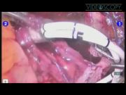
Robotic radical cystectomy and intracorporeal Studer-pouch construction were planned on a 73-year-old man with muscle-invasive bladder cancer. Having completed the uretroileal anastomosis, the Foley catheter with a silk tie at its end is completely withdrawn out through the urethra and replaced with a 20 F Nelaton catheter lumen of which can accommodate two 6F DJSs and a guide wire. The proximal end of the silk tie is grabbed with the forceps and pulled up bringing the newly tied Nelaton tube to the level of urethroileal anastomosis. Spatulated Studer-pouch construction is performed intracorporeally, including Wallace-type ureteroureterostomy. The distal ends of both the ureters are sutured to each other to form a single common duct. Later, the free end of the spatulated left ureter is anastomosed to the proximal end of the ileum. The only remaining opening is between the free spatulated end of the right ureter and the ileum. Nelaton tube is pulled up through the chimney part of the ileal segment from the silk tie. The open end of the Nelaton tube is grasped, and two DJSs are introduced in through its lumen. A 25–30-cm prolene tie is threaded through the holes of DJSs at the most distal ends. The proximal end of the guide wire is grabbed and led into the ureters and fed up to the renal pelves. Free ties hanging from the distal ends of both DJSs are tied to each other and to the indwelling Foley catheter with a slit at its tip. The Nelaton tube is removed out of the urethra, and the distal end of the recently placed guide wire is passed inside the lumen of the Foley catheter backward, which is then slided in until its bifurcation.
Result:
Recovery was uneventful, and the patient was discharged on the seventh postoperative day. Pathologic examination revealed high-grade muscle-invasive transitional cell carcinoma with negative lymph nodes and clear surgical margins. On the postoperative day 21, a pouchogram was obtained, and the Foley catheter and both the ureteral stents were removed altogether. The patient is well and with no evidence of disease 1 year postoperatively.
Conclusion:
We believe that using internal stenting not only prevents intestinal looping around the bridge that would otherwise be formed between the abdominal wall and the intestinal segment, but may also help thinning of the intestinal mucus and prevent clogging of the Foley catheter due to continuous low-pressure irrigation with patient's own urine diverted to the newly formed pouch.
No competing financial interests exist.
Runtime of video: 9 mins 50 secs


Keywords
Get full access to this article
View all access options for this article.
References
Supplementary Material
Please find the following supplemental material available below.
For Open Access articles published under a Creative Commons License, all supplemental material carries the same license as the article it is associated with.
For non-Open Access articles published, all supplemental material carries a non-exclusive license, and permission requests for re-use of supplemental material or any part of supplemental material shall be sent directly to the copyright owner as specified in the copyright notice associated with the article.