
Abstract
Introduction:
Presacral neoplasms are rarely encountered in clinical practice and may present with back pain or symptoms secondary to compression of nerve roots. Complete surgical excision is the treatment of choice and has been attempted through abdominal, abdominosacral, or purely sacral approaches. In selected scenarios, a minimally invasive approach has also been attempted for extirpation of such lesions. 1 We demonstrate an operative video on a transperitoneal laparoscopic approach for extirpation of a presacral neoplasm.
Methods:
A 39-year-old man presented with backache since 6 months. Clinical and laboratory evaluation was normal. Computed tomography urogram revealed a neoplasm located anterior to sacral promontory. The margins were well defined. We decided to embark on a laparoscopic approach for tumor extirpation. Patient was positioned in steep Trendelenberg decubitus. Four ports were employed—one 10-mm camera port at the level of umbilicus, one 10-mm and one 5-mm working port at right and left iliac fossa, and one 5-mm port was inserted at suprapubic area for retraction. The colon was displaced cranially. The peritoneum overlying the tumor was incised. A well-defined plane of the cleavage was identifiable, and dissection was carried out all along following this plane. At the deeper plane, the dissection was carried out meticulously to avoid injury to any neighboring structures. On the posterolateral aspect, a vascular pedicle was identified, which was clipped and divided. Few minor bleeding vessels were detected at the tumor bed that was fulgurated using laparoscopic spatula and monopolar electrocautery. We refrain from over vigorous usage of thermal energy. The neoplasm was completely freed of all surrounding attachments and retrieved in a retrieval bag. Hemostasis was ensured. Drain placement and port closures were undertaken.
Results and Discussion:
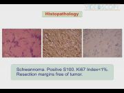
The procedure was completed uneventfully. Operation duration was 130 minutes. Blood loss was 50 mL. The patient made an uneventful recovery postprocedure. Histopathology revealed schwannoma with no evidence of malignant cells. Surgical resection margins were negative. Immunohistochemistry revealed positive S100 and a low proliferative index, indicating very low risk for recurrence. Till last follow-up, the patient is asymptomatic with no sensory or motor neurodeficits and no evidence of recurrent pathology. Laparoscopic extirpation of presacral tumor has been scarcely reported. This modality may be attempted in selected scenarios if preoperative imaging demonstrates an encapsulated lesion with a well-defined periphery. Meticulous dissection is mandated to avoid an iatrogenic insult to neighboring nerve roots. Usage of thermal energy during lesional extirpation also should be restricted to avoid the thermal insult to neighboring neurons. The chief attribute of laparoscopic approach is the magnified view that helps in precise enucleation with preservation of neighboring anatomy. Additional benefits are excellent morbidity and durable outcome.
No competing financial interests exist.
Runtime of video: 5 mins 18 secs


Get full access to this article
View all access options for this article.
References
Supplementary Material
Please find the following supplemental material available below.
For Open Access articles published under a Creative Commons License, all supplemental material carries the same license as the article it is associated with.
For non-Open Access articles published, all supplemental material carries a non-exclusive license, and permission requests for re-use of supplemental material or any part of supplemental material shall be sent directly to the copyright owner as specified in the copyright notice associated with the article.