
Abstract
Introduction:
Ureterocele excision is a procedure that is typically performed in an open fashion. The robotic approach has many challenges associated with it, as well as many advantages. This video will demonstrate a robotic intravesical ureterocele excision, highlighting several novel techniques to aid the procedure, including bladder fixation to the rectus muscle and a unique approach to effective and watertight port closure, which can make a robotic approach a feasible, safe, and effective approach to ureterocele excision.
Methods:
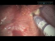
A suitable patient was selected who had a large enough bladder, clinical indication for ureterocele excision, and interest in a robotic approach. The patient had congenital multicystic kidney without much function, and underwent a right nephroureterectomy previously. He had recurrent episodes of hematuria, and a ureterocele was identified via voiding cystourethrogram (VCUG). The ureterocele was a simple orthotopic stenotic ureterocele, seen on VCUG as a filling defect, and confirmed cystoscopically. The decision was made to definitively treat the ureterocele after an attempt at cystoscopic de-roofing failed to relieve the symptoms. After initial cystoscopy was performed, two stay sutures were placed through the rectus muscle and all bladder layers to hitch the bladder to the abdominal wall during the procedure. This prevented the bladder from falling away from the abdominal wall during surgery, and pulling out the robotic ports, and was performed under cystoscopic observation. The robotic ports were placed, along with a Veress needle. The bladder was drained of urine and insufflated, and the camera and instruments placed. The ureterocele was identified, and excised with monopolar scissors. The defect in the detrusor muscle was closed in two layers with vicryl and chromic gut sutures. This patient had a previous nephroureterectomy on the ipsilateral side; therefore, no re-implant was required after the excision. The ports were removed, and the port holes closed using a suture suspension disc. This disc was used as a guide before port placement, and acts as a stencil for proper port and suture placement, ensuring that the preplaced sutures are in the correct position to be able to tightly close the port site.
Results:
This patient did very well postoperatively, with minimal pain and quick return to eating and drinking and ambulating, and was discharged from the hospital 24 hours after his procedure. He had no further hematuria or obstructive symptoms on repeat follow-up examinations.
Conclusions:
With the techniques detailed in this video, the difficulties of robotic ureterocele excision can be easily overcome, allowing the benefits of the minimally invasive approach to be applied to intravesical ureterocele excision. Robotic intravesical ureterocele excision is a safe and effective procedure.
The authors have no professional or financial conflicts of interest to disclose regarding this video or procedure.
Runtime of video: 7 mins 03 secs


Get full access to this article
View all access options for this article.
Supplementary Material
Please find the following supplemental material available below.
For Open Access articles published under a Creative Commons License, all supplemental material carries the same license as the article it is associated with.
For non-Open Access articles published, all supplemental material carries a non-exclusive license, and permission requests for re-use of supplemental material or any part of supplemental material shall be sent directly to the copyright owner as specified in the copyright notice associated with the article.