
Abstract
Introduction:
There is an increasing discrepancy between the number of patients awaiting transplantation and the number of available transplantable organs. To increase the donor pool, live donor kidney transplantation is proving to be an excellent option. Donor nephrectomy for transplantation has become safer over the years and, in experienced hands, is associated with a shorter learning curve. Traditionally, laparoscopic donor nephrectomy has been performed with at least three incisions and a suprapubic one to pass the staplers and use a hand (hand assisted) or an endobag to deliver the kidney. 1 –3 Potential clinical advantages have led to this development, as well as patient's preference for cosmetically superior surgical solutions. 4 Conventional laparoscopy is currently the preferred method for donor nephrectomies. 5,6 The ease and accessibility of less invasive and cosmetically superior methods leads to early hospital discharge, minimal postoperative pain, and a quicker recovery. This encourages more prospective donors to come forward for donor evaluation and donation. 7 Umbilical single-incision donor nephrectomy has been previously described, but a larger umbilical incision might lead to an increased rate of incisional hernias, visible scar, and umbilical deformation because of scaring. 8 –10 This report describes a new method that we have developed for suprapubic single-incision donor nephrectomy under visual control of a 5-mm port (Visiport; Ethicon Endosurgery, Cincinatti, OH) through the umbilicus.
Materials and Methods:
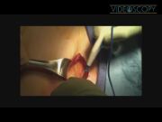
A 54-year-old woman with a history of two prior Caesarean sections underwent suprapubic laparoscopic donor nephrectomy. A 5-mm incision was placed in the depth of the umbilicus. A Veress needle was introduced into the abdominal cavity and pneumoperitoneum created to a pressure of 14 mm Hg. Next, a 6-cm skin incision was made in the existing Pfannenstiehl (suprapubic) scar. Subcutaneous dissection was done to create a pocket by mobilizing the subcutaneous tissue. A 5-mm trocar (Visiport; Ethicon Endosurgery) was placed in the lower and a 12-mm trocar (Visiport; Ethicon Endosurgery) in the upper corner of the incision under direct observation using the umbilical port. Then, the kidney and ureter were mobilized laparoscopically and the ureter was stapled across. A 15-mm port (Ethicon Endosurgery) was placed in the center of the suprapubic incision to introduce a 15-mm Endobag. The kidney was placed in the bag and the remaining attachments of the lienorenal ligament at the upper pole of the kidney were divided using the harmonic scalpel. The patient was administered 2000 units of heparin and the renal artery was stapled across 3 minutes after heparin administration. This was followed by reversal of heparin with 20 units of protamine and stapling off of the renal vein. The suprapubic ports were removed and the incisions in the fascia were joined to remove the kidney. The suprapubic incision was then closed in single layer with No. 1 Maxon. The recipient of this kidney underwent straightforward transplantation and has excellent graft function.
Results:
Suprapubic single-incision laparoscopic donor nephrectomy was successful without any intraoperative or postoperative complications. OR time was 150 minutes with 100 mL of blood loss, and the patient was discharged on the second postoperative day.
Discussion:
Suprapubic laparscopic donor nephrectomy is feasible. This approach is less traumatizing and cosmetically superior compared with conventional laparoscopic and umbilical single-incision donor nephrectomy. However, early mobilization of the upper pole of the kidney is difficult with this approach and the authors have circumvented this problem by dividing these attachments after bagging the kidney and using the endobag as a retracter as well. We believe that this approach is safe and minimally invasive and allows for removal of the donor kidney without incurring undue warm ischemia. The report does, however, have some shortcomings. First, the authors did not conduct a study comparing the two techniques before adopting this technique for laparoscopic donor nephrectomy. We will be pursuing this and will be comparing the data on operative time, estimated blood loss, warm ischemia time, total incision length, peripoerative pain scores, graft outcomes, and complications in these patients prospectively and will be comparing these with the archived data on laparoscopic donor nephrectomies performed using the standard four-port and suprapubic incision technique. Second, for patients with larger body habitus the authors have used bariatric-length instruments, harmonic scalpel, and long suction-tip irrigators. For right-sided donor nephrectomies, the authors have occasionally used another 5-mm port to retract the right lobe of the liver. Third, the authors use this approach with an open mind realizing that donor safety and quality of the graft is of prime importance. In situations where dissection is difficult (for instance, obese donors with a large amount of perirenal fat) the authors place an extra port or extend the Pfannensteil incision to facilitate dissection. Further research will determine whether this approach proves to be safe and results in better donor outcomes as evidenced by pain, cosmesis, and wound-related problems. It will be interesting to find out if it also results in statistically improved graft outcomes compared with the standard laparoscopic nephrectomy and other alternative approaches like robotic live donor nephrectomy.
No competing financial interests exist.
Runtime of video: 7 mins 57 secs


Keywords
Get full access to this article
View all access options for this article.
References
Supplementary Material
Please find the following supplemental material available below.
For Open Access articles published under a Creative Commons License, all supplemental material carries the same license as the article it is associated with.
For non-Open Access articles published, all supplemental material carries a non-exclusive license, and permission requests for re-use of supplemental material or any part of supplemental material shall be sent directly to the copyright owner as specified in the copyright notice associated with the article.