
Abstract
Clinical History:
A 12-month child (10 kg) with ureteropelvic junction obstruction required ureteral stent placement during a robotic pyeloplasty. There was no history of ureteral instrumentation. We describe a novel technique for ureteral orifice dilatation in young children who cannot accommodate traditional ureteral dilators.
Physical Examination:
The patient had normal external genitalia. A 10F rigid cystoscope was inserted into the bladder and the ureteral orifice was located. There were no abnormalities noted within the bladder.
Diagnosis:
A 0.018 glide wire was passed through the ureteral orifice without resistance. A 4.8F ureteral stent would not pass beyond the ureteral orifice. Ureteral stent placement in infants can be challenging because of the small caliber of the orifice and lack of ureteral dilators smaller than 6F. This is typically discovered at time of surgery.
Intervention:
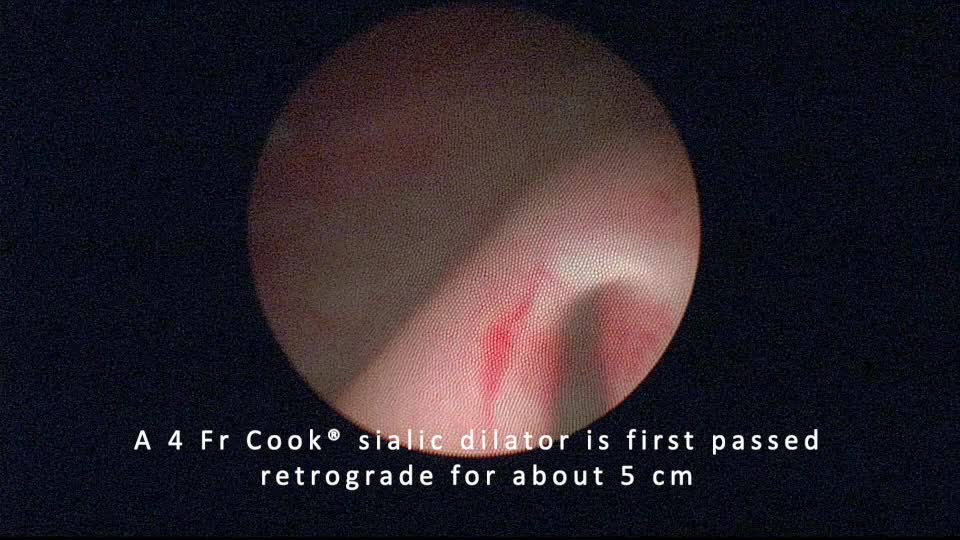
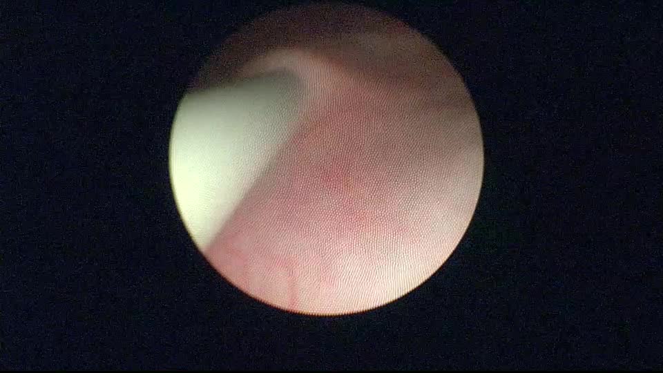
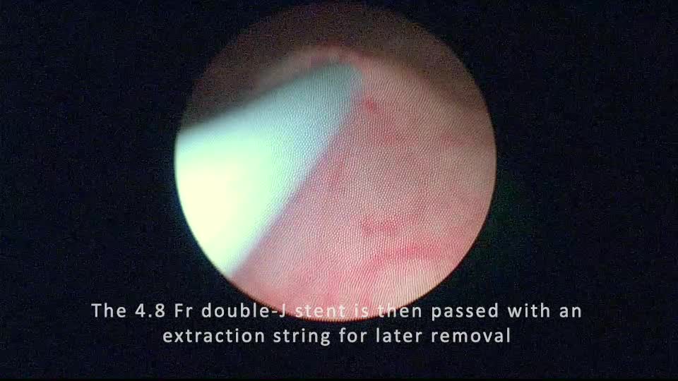
This technique requires the Cook salivary access dilator set, a 0.018 straight glide wire, and a rigid cystoscope. The salivary duct dilators were sequentially passed over the wire starting with the 4F dilator followed by 5F and 6F dilators. This was performed alongside a cystoscope with direct observation for educational purposes but can also be performed under fluoroscopic guidance. Finally, a ureteral stent was advanced with ease.
Follow-Up/Outcomes:
The patient has done well with his ureteropelvic junction repair. Salivary duct dilators offer a smaller caliber dilator that can facilitate retrograde stent placement in infants. We have used this technique with success in several infants whose ureteral orifice would not accommodate a 4.8F ureteral stent.
Authors have received and archived patient consent for video recording/publication in advance of video recording of procedure.
No competing financial interests exist.
Runtime of video: 2 mins 11 secs
Get full access to this article
View all access options for this article.