
Abstract
Introduction:
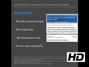
Ureterocele endoscopic treatment became popular because of its lower morbidity compared with open surgery. 1 Endoscopic decompression includes several techniques (U-incision, roof incision, or punctures) and energy sources (Collins knife, diathermic electrocautery, and laser). An electrosurgical incision is the standard endoscopic technique with high decompression rates, however, de novo vesicoureteral reflux (VUR) may occur in up to 62%. 1,2 The watering-can puncture in the bottom of the ureterocele significantly minimizes the risk of secondary VUR, because the collapsed wall acts as an antireflux flap valve. 1 –4 Compared with diathermy electric cautery, laser energy has the characteristic of vaporizing tissue with high precision and less surrounding cell damage or thermal effects. Therefore, compared with traditional electric incision, the possibility of scarring and resealing is less obvious with laser. 1 The aim of this video is to show how to perform the watering-can laser puncture technique for treating ureterocele.
Materials and Methods:
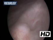
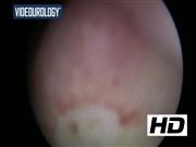
An 11-month male with a history of three urinary tract infections and growth impairment is presented. Renal ultrasonography identified a left duplex kidney with upper pole dilation and bladder scan showed a left ureterocele. Renal scintigraphy showed a functionally impaired left kidney upper pole and cystourethrogram did not show VUR or other abnormalities. Therefore, it was decided to perform an endoscopic decompression of the ureterocele and circumcision. Patient received antibiotics and was placed in a lithotomy position under general anesthesia. A 9.5F cystoscope with a 6F working channel and warm saline was selected. Fluid intake was regulated to the bladder half capacity to promote ureterocele maximum distention. Stabilizing the cystoscope can be challenging since ureterocele may become close to the bladder neck. Therefore, laser fiber must be firmly positioned, otherwise it may recoil and damage the cystoscope. Ureterocele wall may be thick, thus we recommend to push the laser fiber gently to pass through the tissue. In total, 5 to 10 punctures in the bottom of the ureterocele were sufficient to achieve decompression using a 272 µm Holmium YAG laser fiber with 6 Hz and 0.6 J setting.
Results:
Endoscopic procedure lasted 8 minutes, the patient received a Foley catether for 24 hours, and was discharged the next day. Prophylactic antibiotics were discontinued after control ultrasonography with resolution of high-grade hydronephrosis in the third month after the procedure. One-year follow-up showed improved renal function, mild residual hydronephrosis, normal growth, and no infection.
Conclusion:
Endoscopic approach of ureterocele using the watering-can technique is an effective method to achieve decompression. It is a fast and easily reproducible procedure with short hospital stay and reduced postoperative VUR rates.
No competing financial interests exist.
Runtime of video: 4 mins 46 secs
Funding Source: The authors confirm that there are no funding sources in this study or in the writing of the article.
Ethical Approval: The authors confirm that this study has been approved by our institutional board and parents of the patient have consented to publish this article. The authors have received and archived patient consent for video recording/publication in advance of video recording of the procedure. Also, this article is in accordance with the Declaration of Helsinki as revised in 2013.
Get full access to this article
View all access options for this article.