
Abstract
Introduction:
Fibroepithelial polyps (FEP) are rare benign lesions of mesodermal origin that can arise all along the urinary system. 1 –7 They may cause intrinsic ureteropelvic junction (UPJ) obstruction in children and recent literature accounts up to 5% of cases. 8 –15 Symptoms are mainly hematuria and flank pain secondary to the obstruction. 16 –20 The preoperative diagnosis can be challenging as they are difficult to detect by ultrasonography (US). They can be assessed by a radiolucent filling defect in intravenous pyelography or CT scan with contrast. 21,22 The management has not been clearly defined yet, although recent literature suggested an algorithm for diagnosis and treatment of FEP in children. 23 –32
Material, Patients, and Methods:
We retrospectively reviewed two patients with ureteropelvic junction obstruction by FEP treated in our institution.
Results:
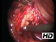
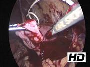
The first case was a 9-year-old boy who presented left flank pain. Repeated US revealed hydronephrosis with a renal pelvis anteroposterior diameter (APD) varying from 10 to 19 mm. There was no evidence of stones on contrast CT scan. An MRI was performed and considered normal at the time. Two years later, as the boy experienced chronic left flank pain, we reviewed the imaging studies and noticed a filling defect on the MRI. The diagnosis was confirmed by intravenous urography showing multiple polyps. Ureteroscopy with yttrium aluminum garnet laser treatment was done twice. Unfortunately, 6 months later recurrent left flank pain occurred and an ureteral stricture was diagnosed. The obstruction was probably a consequence of laser. It was treated by laparoscopic pyeloplasty. We have now 7 years of follow-up and the postoperative course was uneventful. The child is healthy and asymptomatic. The second case was an 11-year-old-boy who experienced two severe left renal colics. The US showed a nondilated renal pelvis with an APD of 6 mm and the enhanced CT scan identified a filling defect suggesting FEP. Ureteroscopy confirmed the presence of multiple FEP obstructing the proximal portion of the ureter. We performed a laparoscopic pyeloplasty during the same procedure. With 2 years of follow-up, the patient is currently doing well without evidence of recurrence based on monitoring for pain and hydronephrosis.
Conclusion:
These cases highlight the difficulty to diagnose UPJ obstructions caused by FEP. We performed ureteroscopy for polyp mapping when filling defect was encountered. Li et al. 8 developed a treatment algorithm dictated by the endoscopic appearance of the ureteral polyp: endoscopic treatment is preferred for one or two pedunculated polyps, whereas pyeloplasty is suggested for multiple polyps to avoid ureteral injury and postoperative ureteral stricture.
Author(s) have received and archived patient consent for video recording/publication in advance of video recording of procedure.
No competing financial interests exist
.
Runtime of video: 8 mins 8 secs
Keywords
Get full access to this article
View all access options for this article.