
Abstract
Objective:
To present the results of a case series of six patients with a duplicated collecting system with ectopic vaginal implantation who underwent laparoscopic heminephrectomy and ureterectomy, accompanied by our last case video.
Introduction:
Duplication of the ureters with infrasphincteric vaginal implantation is an uncommon congenital anomaly and a relatively rare disorder in adulthood as a cause of urinary incontinence. Ectopic ureters are frequently seen in association with a dysplastic upper pole renal moiety. Diagnosis is often difficult, and a high index of suspicion is needed for the correct diagnosis.
Methods:
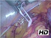
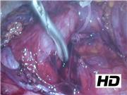
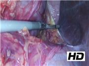
We present a case series of six patients who underwent upper pole heminephrectomy and ectopic ureterectomy in the past 10 years. All surgeries were performed laparoscopically by a single surgeon. The patient's complaints were life-long urinary leakage. All cases underwent magnetic resonance (MR) urography after clinical suspicion. Preoperative CT angiography was routinely performed to understand the anatomy of the renal vasculature. Transperitoneal approach was preferred for all patients. Umbilical camera port, one right-sided instrument port, and one left iliac fossa port were introduced. First, the normal ureter was found, isolated, and secured. Dilated upper pole ureter was found traced through the pelvis and ligated at the level of the cross-over point of the common iliac artery. Then it was traced back and mobilized proximally toward the renal hilum giving extra care not to compromise the blood supply to the normal ureter. If the duplicated upper moiety ectopic ureter was not dilated, it could be difficult to separate from the normal ureter. In such cases, it is important to secure and trace both ureters to be sure to find the correct ureter. It was isolated from the accessory lower pole vasculature and was cut. The remaining pelvis was dissected from the renal pedicle. The right kidney was identified and mobilized. The upper dysplastic pole of the right kidney was located and accessory vessels to the upper pole moiety were clipped. Demarcation was seen between dysplastic tissue and normal renal parenchyma, and heminephrectomy for poorly functioning upper pole moiety was completed with ultrasonic energy.
Results:
All patients were female, and the age ranges were between 19 and 38 years. Duplicated collecting systems and ectopic ureters were on the right side for four patients and on the left side in the other two patients. According to MR urography, the upper pole dysplastic kidneys were prominently dilated in five patients, in the remaining one patient, the upper pole was not dilated. All surgeries were completed laparoscopically, and mean hospital stay was 3 days. There was no postoperative early complication in all cases. All patients were fully continent after catheter removal on postoperative day 1.
Conclusions:
Ectopic ureteral duplication should be considered in the differential diagnosis of young women presenting with life-long wetting. The best radiologic imaging modality in the diagnosis of this rare disease is MR urography. The surgical approach depends on the surgeon's experience. However, considering the benefits of laparoscopy, it should be the preferred approach over the open surgery for the management of patients with duplicated collecting system with ectopic implantation.
No competing financial interests exist.
Run time of video: 4 mins 55 secs
Get full access to this article
View all access options for this article.