
Abstract
Introduction and Objectives:
Endoscopic enucleation of the prostate (EEP) is an established, safe, and effective alternative to transurethral resection of the prostate and open prostatectomy for the treatment of lower urinary tract symptoms secondary to benign prostatic obstruction. All EEP procedures, regardless of the source of energy used (holmium, thulium, green laser, and bipolar energy), require a steep learning curve (LC) and during this LC the risk of stress urinary incontinence (SUI) could be increased. It is very important to know the surgical anatomy of the male external urethral sphincter (EUS) well to avoid SUI. The morphology of the male EUS can be assessed by various types of diagnostic imaging: MRI and transrectal ultrasonography (TRUS).
Objectives:
(1) To review the surgical anatomy of the male EUS by microscopic tissue sections of cadaveric specimens and by MRI and TRUS images. (2) To evaluate the intraoperative TRUS images of the male EUS during laser EEP and to report the morphology before and after surgery.
Materials and Methods:
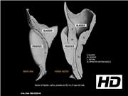
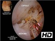
We reported a summary of the topographic anatomy of the male EUS based on microscopic and macroscopic tissue sections from male cadaveric specimens, TRUS images, and MRIs. Eleven patients underwent EEP, five of whom underwent holmium laser enucleation of the prostate (HoLEP) and six underwent green laser enucleation of the prostate (GreenLEP). All the procedures (HoLEP and GreenLEP) were done with the “en bloc” (on lobe) technique (with early sphincter separation, avoiding mechanical and thermal injury of the EUS). All patients' EUS was studied by intraoperative TRUS using a linear probe before and after surgery. However, in this video, only the results of one case have been presented. Morphologic parameters of the EUS (shape, length of echo pattern, and thickness) were evaluated. In the postoperative follow-up appointments at 1, 3, and 6 months, the International Prostate Symptom Score, quality of life, maximum urinary flow rate, post-void residual volume, prostate specific antigen levels, International Index of Erectile Function, and SUI were assessed.
Results:
A detailed topographic anatomical study of the male EUS has been presented. The male EUS was easily detectable by TRUS as a hypoechoic circular shaped structure surrounding the membranous urethra, which remained unchanged before and after the EEP procedure. The patient was discharged 24 hours postoperation without complications. The bladder catheter was removed at 24 hours after surgery and 3 months later all the parameters showed significant improvement. There was no SUI as a result of the procedure.
Conclusions:
First and foremost, it is imperative that the surgeon has a complete understanding of the EUS surgical anatomy before performing EEP to avoid SUI during the LC. As demonstrated previously in the literature, EEP represents an established safe and effective technique for the complete removal of the adenomatous prostate tissue. This is not an easy procedure and requires a steep LC. During “en bloc” EEP, carefully dissecting the apical lobes from the EUS is fundamental in preventing SUI, as it protects the sphincter from thermal and mechanical dissection damage.
No competing financial interests exist.
Runtime of video: 7 mins 51 secs
Another version of this video was presented in the 2018 Annual European Association of Urology (EAU) Congress in Copenhagen, Denmark.
Get full access to this article
View all access options for this article.