
Abstract
Introduction:
In transvesical natural orifice transluminal endoscopic surgery (NOTES), 1,2 techniques of endoscopic closure of the bladder perforation have been reported, to avoid or to decrease the bladder drainage time. For percutaneous endopyeloplasty, we perform endoscopic sutures using a needle holder through the nephroscope and a conventional suture. 3,4 We evaluate the feasibility of endoscopic bladder suturing using the same technique. We present our initial experience of endoscopic bladder suturing during pluck transurethral detachment of the intramural ureter, in the first part of nephroureterectomy.
Materials and Methods:
We present the feasibility of endoscopic bladder suturing in a 70-year-old woman, with transitional cell carcinoma of the left renal pelvis and upper calyxes, diagnosed by ultrasonography and a CT scan for gross hematuria. Retrograde flexible ureteroscopy and biopsy confirmed the carcinoma. The study was approved by the hospital ethics committee. After informed consent, the patient underwent an endoscopic closing of the ureteral orifice before the intramural ureter is detached in the first part of nephroureterectomy. Then, the bladder is sutured after ureteral detachment.
Operative Technique:
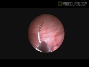
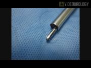
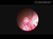
Under general anesthesia, the patient is placed in the lithotomy position. A 26F Amplatz sheath is placed in the bladder via the urethra. Cystoscopy is performed using a 24F nephroscope. The nephroscope is retrieved, and a lengthened 3.5-mm pediatric laparoscopic needle holder is placed through its working port. A conventional 13-mm needle, of an absorbable suture, is grasped in the middle by the needle holder in the conventional manner. The nephroscope is introduced in the bladder with the suture dragged alongside the nephroscope. The ureteral orifice is closed in a conventional suturing manner using a figure-of-eight suture. The nephroscope is retrieved, and the resectoscope is introduced. The bladder is washed thoroughly to evacuate the possible tumor cells. The ureteral orifice is detached using a Collins knife electrode. The resectoscope is removed, and the nephroscope is once again inserted. The bladder wound is sutured using two figure-of-eight sutures. A 16F Foley catheter is inserted in the bladder. Then, open nephroureterectomy is performed.
Results:
The endoscopic suturing of the ureteral orifice and the bladder was successfully completed in the first attempt. The time of ureteral suturing was 9 minutes, and the time of bladder suturing was 22 minutes. The Foley catheter was removed in the third postoperative day. No perioperative complication was noted.
Conclusions:
The endoscopic suture of the bladder was possible, and allowed a short-bladder catheterization, which suggests the feasibility of the endoscopic bladder suture using a needle holder through the nephroscope and a conventional suture. The endoscopic suture might be used for transvesical NOTES, in case of bladder perforation or traumatic rupture. In addition, it might have further hypothetical applications: removal and neck closure of bladder diverticulum; endoscopic treatment of retrovesical hydatid cyst 5 ; Heineke-Mikulicz plasty of short urethral strictures, bladder neck contracture, and vesicourethral or pouch-urethral anastomosis stenosis; realignment and suture of urethral rupture; or endoscopic ureteral reimplantation and vesicourethral reflux treatment. However, further technical experience and more studies are necessary to establish endoscopic suturing applications and effectiveness.
The authors have no conflicts of interest or financial ties to disclose.
Runtime of video: 8 mins
Keywords
Get full access to this article
View all access options for this article.