
Abstract
Introduction/Aims of Study:
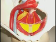
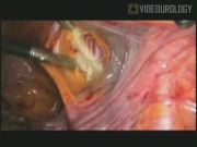
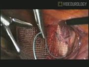
Major difficulties of sacral colpopexy are ileus and defecation difficulties caused by less space in the lower pelvis. The iliopectineal ligament has been used over a long period for Burch operation and the pectopexy does this either. The lateral positioning of the mesh beneath the round ligament has enabled a sufficient fixation without restriction of the bowel. The study shows the good postoperative outcome of the technique. Multiple publications show that for the reconstitution of a physiological axis of the vagina regarding size, depth, and slant, a sacropexy seems to be the most adequate approach. 1 –5 For more than 6 years we have been performing laparoscopic sacral colpopexy in the treatment of vaginal vault prolapse. Our cure rate of 92.1% 6 and our experience of >600 procedures led our view to some weak points of the technique and encouraged us in developing a new technique. As obesity is one of the major risks for vault prolapse, it can be a challenge for surgery. The sigmoid colon is often enlarged by fatty tissue. In this case there is less space for the placement of a mesh between the vagina and the sacrum. Consecutively, pain or defecation problems can result. The pectopexy uses the iliopectineal ligament on both sides for the mesh fixation, 7 so there is no restriction caused by mesh. We performed this technique since 2007 in cases of less space in the pelvis (especially obese women) and difficulties caused by former surgery (93 cases). We saw one relapse caused by an insufficient stump-fixation. No major complications caused by surgery were seen. There was one revision surgery after 1 year caused by an enterocele. So we had no occasion for additional treatment of the “cul-de-sac” routinely. With our trial we want to show that the new technique is safely performable like sacropexy and shows comparable results in long term.
Materials and Methods:
A randomized trial was started in October 2010 to compare the gold-standard laparoscopic sacral colpopexy to the pectopexy. For evaluating the long-term data, we use the International Consultation on Incontinence Questionnaire-Vaginal Symptoms (ICIQ-VS) 8 and a standardized documentation to acquire data, including the pelvic organ prolapse quantification (POP-Q), for all segments. Only defects POP-Q II and higher were included. Follow-up examination would start 1 year after surgery. We documented the operation time, blood loss, body measurements, and different complications for the postoperative outcome.
Results:
The postoperative data of the first 72 patients were evaluated and showed no difference in complication rate or hospital stay (4–5 days). No major complications (bowel injury, ileus, or mesh infection) were seen in either group. We saw one urinary infection in the pectopexy group. No defecation problems or denovo incontinence was found. The mean operation time was 44.57 minutes for the pectopexy and 52.75 minutes for the sacral colpopexy. Blood loss was documented with 4.8–14.75 mL. The international interest and the prize we received for the video encouraged us to publish the video with the postoperative data.
Discussion:
The first data show that the new technique carries no new risks and can be performed as same as the classic gold standard, the sacral colpopexy. We regard the pectopexy as an enrichment of our surgical portfolio in prolaps surgery. Only the long-term data can show if the procedure is performable routinely.
No competing financial interests exist.
Runtime of video: 7 mins 41 secs
The video shows the technique we first published in October 2010. 7 The video was shown on the international conference of the European Society of Gynaecological Endoscopy (ESGE) in London (9/2011). The author received the Lilo Mettler award for the best video presentation.
Get full access to this article
View all access options for this article.