
Abstract
Abstract
Introduction:
Nowadays, the standard procedures of removing benign adrenal tumors are minimally invasive surgical techniques using the laparoscopic or the retroperitoneoscopic route. In adrenal carcinomas most surgeons still prefer open accesses. Some adrenal carcinomas create a tumor thrombus (TT) in big veins. We present the case of adrenal cancer with a TT in the inferior vena cava (IVC) that was removed through the retroperitoneoscopic route.
Patient and Method:
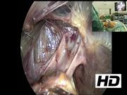
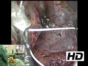
In a 43-year-old female patient a 4 cm mass of the right adrenal gland was incidentally diagnosed. Hormonal tests were negative. Magnet resonance imaging showed a small TT in the IVC. We decided to remove the adrenal tumor and TT by the posterior retroperitoneoscopic approach.1,2 Three ports were introduced underneath the 11th and 12th rib. The retroperitoneal space was opened by meticulous dissection under carbon dioxide pressure of 20 mm Hg. The IVC was circularly dissected and clamped inferiorly and superiorly to the adrenal tumor. After lateral mobilization, the adrenal mass was placed into a plastic bag. IVC was opened by circular excision of the junction of the adrenal vein. By this maneuver the TT was mobilized to glide into the bag. Fragmentation of tumor tissue and tumor cell spillage could be avoided. The defect of the IVC was closed by running suture. After declamping of the IVC, free blood flow was maintained. The tumor was removed by fragmentation out of the bag. No drain was used.
Results:
Operating time was 170 minutes. Blood loss was neglectable. Mobilization and food intake started on day of surgery. The postoperative course was uneventful. Histopathology report described an adrenocortical cancer with confluenting necroses and a Ki-67 proliferation rate of 15%. Postoperatively, adjuvant mitotane therapy was started. After a follow-up of 18 months, patient is disease free without any evidence of local or distant recurrence.
Summary:
This video shows—for the first time—an oncologic minimally invasive extirpation of adrenal carcinoma with a TT in the vena cava. Furthermore, it demonstrates principle and method of safe and bloodless surgery by vessel control, useful in minimally invasive and in open surgery.
No competing financial interests exist.
Authors' Contributions: All three authors participated in collection, selection, and editing of the video material.
Consent: Patient gave written informed consent to show surgery and other pictures, including her body and face.
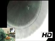
Runtime of video: 11 mins 57 secs
Get full access to this article
View all access options for this article.