
Abstract
Introduction:
Ansa-to-recurrent laryngeal nerve (ANSA-RLN) anastomosis procedures in children with unilateral vocal fold immobility (UVFI) are considered a first-line treatment strategy for long-standing immobility. Pediatric UVFI is often caused by iatrogenic injury (patent ductus arteriosis [PDA] ligation, thyroidectomy, etc.) in which the status of the recurrent laryngeal and vagal neural circuit is often unknown. Understanding the neural circuitry from the vagus to RLN is an important prognostic factor in ANSA-RLN voice outcomes. The following video article describes through detailed high-yield surgical videos the steps necessary to perform pediatric ANSA-RLN aided by vagal nerve evoked electromyography (EMG). Patient outcomes are better with intact RLN-vocal cord circuitry and, therefore, patients will have similar outcomes, regardless of the timing of their surgery after UVFI, as long as the evoked EMG is intact. Should evoked EMG suggest the RLN circuit is not intact, alternative surgical approaches may be utilized (injection medialization laryngoplasty and open medialization laryngoplasty).
Surgical Procedure:
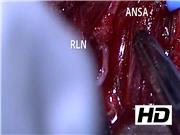
Electromyographic hookwire electrodes (Medtronic, Jacksonville, FL) are inserted into the bilateral thyroarytenoid and posterior cricoarytenoid muscles under direct laryngoscopy. Awake bilateral intraoperative spontaneous EMG is then completed before the open aspect of the procedure. Should the surgeon wish to only perform intra-operative evoked EMG, electrodes are only necessary on the ipsilateral vocal cord. The patient is then taken out of suspension and the neck is prepped and draped in the standard manner. It is important not to use paralysis during the case as it will interfere with the ability to conduct accurate intraoperative nerve monitoring. Contrary to previously described ANSA-RLN cases, it is necessary to identify and expose the vagus nerve. Before dissection of the ANSA and proceeding with ASNA-RLN anastomosis, the surgeon now performs evoked EMG. If the vagal-RLN connection is deemed intact, the surgeon will proceed with the surgery. The ansa cervicalis is then dissected in its entirety, over the carotid sheath, until it loops back along the jugular vein. Before transecting the ansa, the RLN is then identified and dissected. Enough length is needed to allow the RLN to be turned superiorly to achieve a tension-free anastomosis with the ansa cervicalis. As many patients have only a short stump of RLN at the cricothyroid insertion point, it is imperative to dissect enough length of the ansa to ensure tension-free anastomosis. At this point, the ansa can be transected with a microscissor and tunneled under the straps to bring it to the transected RLN. Using either loupe or a microscope, an epineural-to-epineural anastomosis is achieved with at least two 9-0 nylon sutures. A drop of fibrin can be placed over the anastomosis before closing the neck over a rubber band drain. Before waking up, the child receives an injection medialization laryngoplasty as the ANSA-RLN anastomosis typically takes 6 to 9 months to achieve its affect. This approach is similar regardless of the type of iatrogenic injury (thyroidectomy, PDA ligation, etc.).
Conclusion:
The ANSA-RLN anastomosis with adjunct evoked EMG is a readily available technique with limited added morbidity.
No competing financial interests exist.
Runtime of video: 6 mins 47 secs
Setup supplementary video:
The setup of the machine, including creation of a custom settings tab, for the ANSA-RLN procedure is described in detail in this supplemental video: (https://vimeo.com/377603129).
Get full access to this article
View all access options for this article.