
Abstract
Abstract
Introduction:
Craniopharyngiomas represent <1% of all primary central nervous system tumors but are the most common intracranial nonglial tumor in children. 1 Considering tumors of the sellar/suprasellar region, it represents the second most common tumor after pituitary adenoma. Despite the benign histological appearance, it can be a great surgical challenge because of their potentially tendency to tightly adhere to nearby vital structures, including optic apparatus, Circle of Willis vessels, infundibulum, and hypothalamus. Various transcranial and transsphenoidal routes have been described for removal of craniopharyngiomas. Over the past two decades, keyhole surgical approaches have been increasingly used. The two most common approaches are the endonasal endoscopic approach and supraorbital “eyebrow” craniotomy. In our view, the endonasal trajectory is naturally suited for the great majority of craniopharyngiomas situated in the retrochiasmal space and extending into the third ventricle, whereas the supraorbital approach is ideal for tumors in the pre- or suprachiasmatic space or those with significant lateral extension. 2 This video discusses and demonstrates the indications and technical details of the extended endonasal endoscopic transsellar transplanum approach for retrochiasmal craniopharyngiomas.
Materials and Methods:
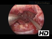
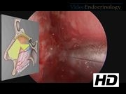
Fully endonasal endoscopic craniopharyngioma surgery at our center is performed utilizing a bimanual-binasal, two surgeon technique. All operations are performed in the endoscopic operating suite, using frameless image-guided navigation and Doppler ultrasound. Zero degree endoscope is used for visualization in most of the procedure. The 30° and 45° angled endoscopes are very useful at various stages particularly for lateral and suprasellar visualization. Typically, an otolaryngologist performs the initial endonasal approach to the sphenoid sinus. Given that a high-flow grade 3 cerebrospinal fluid leak is expected upon tumor removal, a pedicled nasoseptal flap is raised at the beginning of the surgery.3,4 Next, the neurosurgeon, with the otolaryngologist “driving” the endoscope, utilizes a binostril approach to perform the remainder of the sellar and planum exposure, tumor removal, and skull base reconstruction.
Case Example and Results:
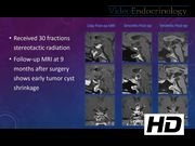
An extended endonasal endoscopic transplanum transsellar craniopharyngioma (papillary subtype) resection is demonstrated in a 54-year-old male who presented with central hypogonadism and borderline hypothyroidism. The patient was strongly in favor of a gland and infundibulum-sparing surgery in hopes of avoiding panhypopituitarism. Hence, the surgical goals were complete removal of the solid tumor portion with the preservation of the pituitary gland and infundibular connection to the hypothalamus. The video shows the endonasal approach, sphenoidotomy, sellar and planum exposure, tumor removal, and skull base reconstruction. As anticipated, near complete total tumor resection was achieved in this patient with complete removal of the solid tumor portion. His initial postoperative MRI shows that the solid contrast-enhancing portion of the tumor has been removed, while the cyst lining and stretched infundibulum is still visualized as is the pituitary. The optic chiasm is well decompressed. The patient has done well since surgery. As planned, he was recently treated with stereotactic radiotherapy (30 fractions) for residual tumor and cyst lining. His hypogonadism and hypothyroidism have persisted, and he developed new persistent diabetes insipidus, despite stalk preservation. He is currently on thyroid, testosterone, and desmopressin. His recent MRI 9 months after surgery and post-stereotactic radiotherapy shows early cyst shrinkage and no solid tumor recurrence.
Conclusions:
The endonasal endoscopic approach has been shown to be safe and effective for retrochiasmatic craniopharyngiomas. This approach obviates brain retraction, minimizes optic apparatus manipulation, and allows early identification of the pituitary gland and infundibulum. A team approach that includes both neurosurgery and otolaryngology is highly recommended for these technically challenging cases.
D.F. Kelly has a royalties agreement with Mizuho, Inc. For all other authors, no competing financial interests exist.
Runtime of video: 11 mins 5 secs
Get full access to this article
View all access options for this article.