
Abstract
The goal for this work was to characterize basic biological properties of a novel Arthrospira platensis-based aqueous cyanophyta extract (ACE), enriched in the known anti-inflammatory cyclooxygenase-2 (COX-2) inhibitor phycocyanin (PC), but also containing a high level of non-PC bioactive compounds. Antioxidant properties were tested in parallel in the Folin–Ciocalteu assay (chemical antioxidant capacity) and in the cellular antioxidant protection (CAP-e) bioassay, where both the PC and the non-PC fractions contributed to the antioxidant capacity and CAP of ACE. In contrast to the COX-2 inhibition seen in the presence of PC, the inhibition of enzymatic activity of the inflammatory mediator Lipoxygenase was associated specifically with the non-PC fraction of ACE. Inhibition of formation of reactive oxygen species (ROS) was evaluated using polymorphonuclear cells from healthy human donors. The inhibition of ROS formation was seen for both the PC and non-PC fractions, with ACE showing the most robust effect. The effects of PC, non-PC, and ACE on clotting and clot lysing was tested using a modified Euglobulin fibrinolytic assay in vitro. In the presence of PC, non-PC, and ACE, the time for clot formation and lysis was not affected; however, the clots were significantly more robust. This effect was statistically significant (p<.05) at doses between 125–500 μg/mL, and returned to baseline at lower doses. Both PC and the non-PC fraction contributed to the antioxidant properties and anti-inflammatory effects, without a negative impact on blood clotting in vitro. This suggests a potential benefit for the consumable ACE extract in assisting the reduction of inflammatory conditions.
Introduction
C
PC, the photoharvesting pigment unique to cyanobacteria, is a water-soluble phycobilin protein capable of exerting potent antioxidant protection and anti-inflammatory activities. 7 –10 PC represents a significant component of blue-green algae, as it can constitute up to 15% of its dry weight when harvested. In addition to the well-documented selective inhibition of cyclooxygenase-2 (COX-2) activity, animal studies of acetic acid-induced colitis in rats found that PC reduced myeloperoxidase (MPO) activity and the corresponding tissue inflammation. 11 In terms of its antioxidant activity, in vitro studies have reported PC scavenging alkoxyl, hydroxyl, and peroxyl radicals, as well as reacting with peroxynitrite and hypochlorous acid, 12 thus reducing cellular oxidative damage.
In addition to the PC component, blue-green algae contains significant amounts of water-soluble phenolic compounds, oligo- and polysaccharides, and PUFAs, many of which are known to exhibit significant antioxidant and anti-inflammatory properties. Polyphenols have been observed to act as free radical scavengers, hydrogen-donating compounds and induce cellular antioxidant defense modulating proteins and gene expression. 13 Specifically, in an evaluation of the antioxidant capacity of Arthrospira maxima extracts, the phenolics found to be responsible for its antioxidant properties were organic acids (including caffeic, chlorogenic, quinic, salicylic, sinapic, and trans-cinnamic acid). 14 Additionally, PUFAs such as gamma-linolenic acid (GLA), an omega-6 fatty acid, play a significant role in the modulation of inflammatory responses through an oxidative metabolism to produce anti-inflammatory eicosanoids (such as prostaglandins). 15
In addition to their fundamental roles in global carbon cycling and nitrogen fixation, cyanobacteria such as Arthrospira platensis and A. maxima have become increasingly important economically in the production of human dietary supplements. Due to their diverse biological activities, in vitro and in vivo studies have been conducted to study selective COX-2 inhibition, 16 antioxidant, 17 and anti-inflammatory activity, 18 as well as neuroprotectant 19 and hepatoprotectant 20 properties. Therefore, due to the well-documented properties of blue-green algae, A. platensis and A. maxima are gaining acceptance as nutritional support in the prevention of various chronic metabolic and inflammatory conditions.
Crude A. platensis powder contains nonwater-soluble components, including chlorophyll, lipids, and potentially inflammatory cell wall components such as glucans and complex glycopolysaccharides. The present research focused on evaluating a novel PC-enriched aqueous cyanophyta extract (ACE), 21 enriched in soluble bioactive components, compared with crude dried Arthrospira. The present article encompasses the in vitro data in three distinct areas: antioxidant properties and bioprotection, anti-inflammatory activity, and blood clotting and fibrinolysis.
Materials and Methods
Reagents
Histopaque 1077 and 1119, RPMI-1640 culture medium,
Product and fractions
The test product used in this study is a proprietary ACE, derived from Arthrospira (subspecies platensis), and commercially available as a dried powder under the name Cyactiv™ (referred to as ACE in this article), produced by a proprietary process involving aqueous extraction.
Pure PC was produced by subjecting the ACE aqueous extract described above to centrifugation, followed by passage over an Amberlite FXP66 resin, followed by chromatography over silica. 22 A PC-free extract of ACE was subsequently produced by eluting the resin with aqueous alcohol (hereafter referred to as the non-PC fraction). Both fractions were freeze dried. In preparation for adding product to laboratory assays, 0.5 g dried powder was added to 5 mL PBS, mixed by inversion and vortexing, and allowed to incubate for 1 h on a rocker. The remaining solids were removed by centrifugation, followed by filtration through a 0.22 μm cellulose acetate filter. Serial dilutions were prepared in PBS.
Inhibition of lipoxygenase enzymatic activity
Testing of the effect of the test products on the enzymatic activity of lipoxygenase was tested using the Lipoxygenase Inhibitor Screening Assay Kit (Cayman Chemical, Ann Arbor, MI, USA), according to the manufacturer's instructions. In brief, purified soybean Lipoxygenase was allowed to react with the substrate arachidonic acid in the absence versus presence of test products. The hydroxyperoxides produced as a result of the Lipoxygenase enzymatic reaction were measured in a colorimetric assay, and absorbance read in a microplate reader (BioTek PowerWave, Winooski, VT, USA) at 490–500 nm absorbance.
Inhibition of COX-2 enzymatic activity
Testing of the effect of the test products on the enzymatic activity of COX-2 was tested using the COX-2 Enzyme Activity Assay Kit (Cayman Chemical), according to the manufacturer's instructions. In brief, arachidonic acid served as a substrate for human recombinant COX-2 enzyme, allowing COX-2 to produce prostaglandin. The assay measures PGF2α produced by SnCl2 reduction of COX-derived PGH2. The PGF2α levels produced in the presence versus absence of test products were quantified through an enzyme immunoassay using an antibody that binds to all major prostaglandin compounds.
Folin–Ciocalteu assay
The antioxidant capacity was evaluated by the Folin–Ciocalteu assay. The Folin–Ciocalteu's phenol reagent was added to serial dilutions of the test products and incubated for 5 min. To start the chemical reaction, sodium carbonate was added, and the reaction allowed to continue for 30 min at 37°C. The optical absorbance at 765 nm was read in a colorimetric plate reader (BioTek PowerWave).
Purification of polymorphonuclear cells and erythrocytes
Healthy human volunteers between the ages of 18 and 65 years served as blood donors after written informed consent was obtained, as approved by the Sky Lakes Medical Center Institutional Review Board (FWA2603). Isolation of polymorphonuclear (PMN) cells was performed as previously described. 23,24 The PMN cells were used for evaluation of anti-inflammatory activity in the assay for production of reactive oxygen species (ROS). The erythrocytes were stored at 4°C in aliquots for later use in the cellular antioxidant protection (CAP-e) bioassay.
Cellular antioxidant protection assay
The CAP assay using erythrocytes (CAP-e) was performed according to the method published by Honzel et al., 25 but using an accelerated and more sensitive microplate-based protocol, where a red blood cell suspension was treated with test products before labeling with a precursor dye as a reporter for oxidative damage, followed by the induction of oxidative stress in the cells using AAPH. Six wells served as negative controls (no oxidative damage was induced) and six wells served as positive controls (maximum oxidative damage in the absence of any antioxidants). Gallic acid was used as a standard reference compound. Serial dilutions of test products were tested in duplicate. The green fluorescence intensity, as a measurement of oxidative damage, was recorded at 488 nm using a Tecan SpectraFluor plate reader (Durham, NC, USA). The CAP was calculated as the inhibition of oxidative damage as indicated by the reduced fluorescence intensity in the wells, where the cells were pretreated with test products, when referenced to the baseline (negative controls), and maximum oxidative damage (positive controls).
Reduced free radical formation by PMN cells
The PMN cells were incubated at 37°C, 5% CO2 for 20 min, either untreated or treated with serial dilutions of test products, as previously described. 23 –25 The PMN cells were washed twice in PBS to remove any unbound and unabsorbed compounds from the test product. The untreated versus product-treated PMN cells were subsequently loaded with a precursor dye DCF-DA, and incubated for 1 h at 37°C to allow the precursor dye to be absorbed into the PMN cells. All samples, except for the triplicate negative control samples, were then exposed to 167 mM H2O2 for 45 min to induce severe oxidative stress. Samples were washed twice in PBS to remove the peroxide, transferred to ice-cold RPMI 1640 medium, and stored on ice in the dark. The DCF-DA fluorescence intensity was immediately analyzed by flow cytometry. Data were collected in triplicate for controls and for each dose of the test products extracts. The mean fluorescence intensity of PMN cells was compared between untreated, H2O2-treated, and product-pretreated cells. A reduction in mean fluorescence intensity in samples pretreated with test products before challenge with H2O2 indicated that a given test product was able to reduce the formation of ROS in PMN cells under conditions of oxidative stress.
Euglobulin assay for clotting and fibrinolysis
Testing of clot formation and fibrinolysis was performed using the Euglobulin assay. Whole blood was drawn into sodium citrate vacutainer vials. Samples of 1140 μL of the whole blood were distributed into glass vials, and 60 μL test product added to each vial. Untreated whole blood served as control. The euglobulin fractions from each vial were obtained using acetic acid. The euglobulin fraction was resuspended in NaCl/NaB3 solution under gentle agitation. Samples were added to a prewarmed round-bottom 96-well microplate. Eight wells were plated from the untreated control samples, and duplicate wells of samples treated with serial dilutions of test products were plated in parallel. Clotting was initiated by the addition of CaCl2. The clot formation and subsequent fibrinolysis was measured by the absorbance at 405 nm using a BioTek PowerWave plate reader, where measurements were taken every 6 min for 16 h.
Statistical analysis
Statistical analysis was performed using Student's two-tailed independent t-test. Levels of statistical significance between data sets were: Significant if the p-value was less than .05 (*), highly significant if p<.01 (**), and very high level of significance if p<.001 (***).
Results
This study compared the PC-enriched cyanophyta extract ACE to both its non-PC fraction and to pure PC. The absence of PC in the non-PC fraction was documented by spectral scanning, showing absence of a peak at the 620 nm wavelength characteristic for PC (Fig. 1).
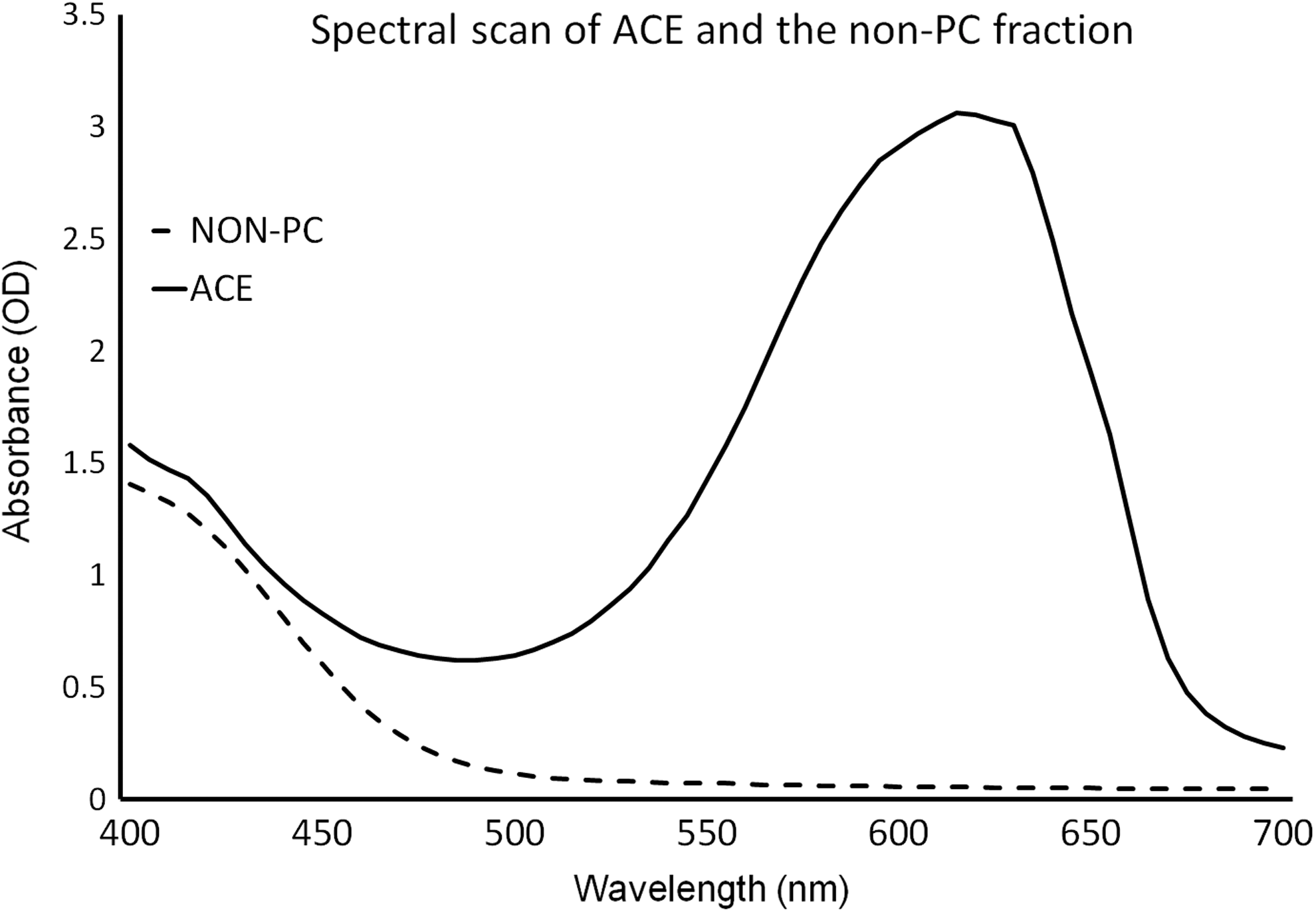
Absorbance spectrum for the aqueous cyanophyta extract (ACE, solid line) and the non-Phycocyanin (non-PC, dashed line) fraction. The robust peak at 620 nm seen in ACE is a characteristic property related to PC content. The absence of the peak at 620 nm in the non-PC fraction is clear. The detection limit for PC at the 620 nm wavelength corresponds to 0.02 μg/mL.
The cyanophyta extract ACE was tested for its ability to inhibit the inflammatory enzymes COX-2 and Lipoxygenase. The ability of the extract to inhibit the enzymatic activity was comparable to the inhibition of Lipoxygenase activity seen for the non-PC fraction of ACE (Fig. 2). While PC is known to possess potent COX-2 inhibitory properties, 16 the inhibition of Lipoxygenase is predominantly caused by non-PC compounds.
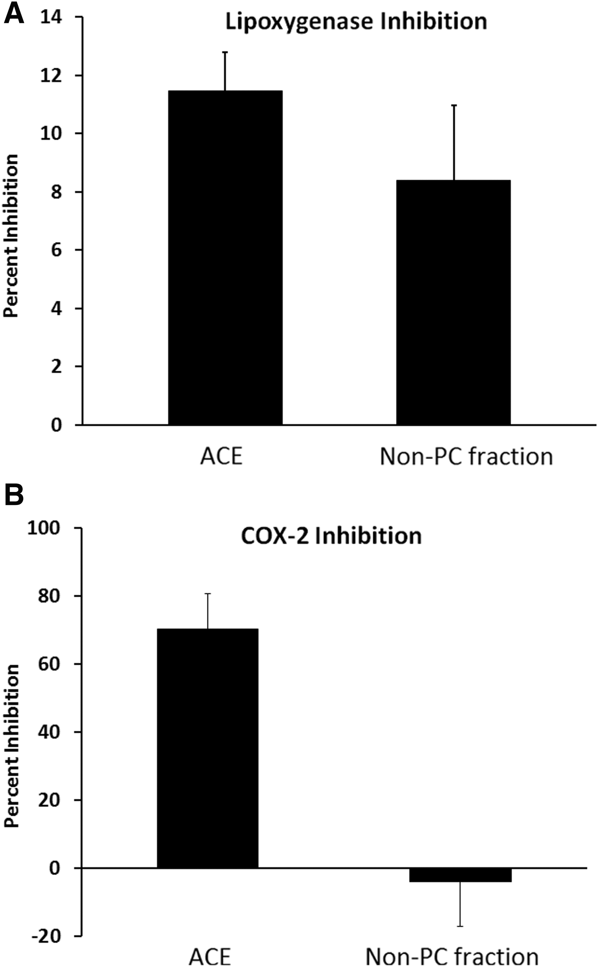
Inhibition of Lipoxygenase and Cyclooxygenase-2 (COX-2) enzymatic activity by ACE at 0.01 mg/mL, compared with the extract at the same dose after removal of PC (non-PC fraction).
The antioxidant capacity of ACE and the two fractions was tested using the Folin–Ciocalteu assay (Fig. 3). The antioxidant capacity of the non-PC fraction exceeded that of PC and ACE; this difference was highly significant across a wide dose range (p<.001). Interestingly, the antioxidant capacity of ACE did exceed that of pure PC, and this was statistically significant at some doses (p<.05). The slightly higher antioxidant capacity of ACE compared with pure PC is likely due to the presence of the antioxidants in non-PC fraction. The ability of the test fractions to provide CAP was tested using the CAP-e bioassay (Fig. 4). The whole ACE product showed higher CAP than either PC or the non-PC fraction. Thus, both the chemical and cellular antioxidant data points to an additive effect between PC and non-PC compounds in ACE.
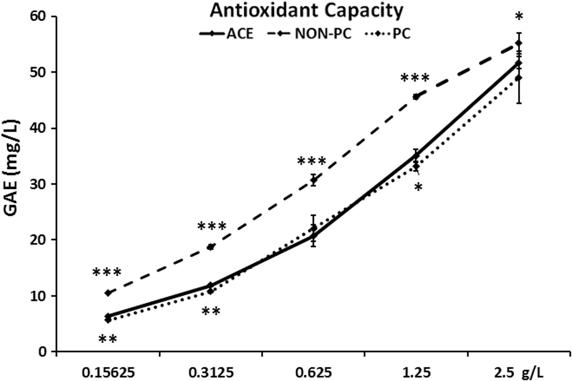
The antioxidant capacity of the PC-enriched ACE is shown compared with the antioxidant capacity of PC and the non-PC fractions of ACE. The higher antioxidant capacity of the non-PC fraction of ACE indicates that compounds other than PC contribute to the overall antioxidant capacity of ACE. For the dose range shown, the antioxidant capacity of non-PC was significantly higher than both ACE and PC (p<.05). The antioxidant capacity of ACE was mildly elevated compared with PC alone; data reached levels of statistical significance for test points with small error bars. Levels of significance are indicated by asterisks: p<.05 indicated by *, p<.01 indicated by **, and very high level of significance if p<.001 is indicated by ***.
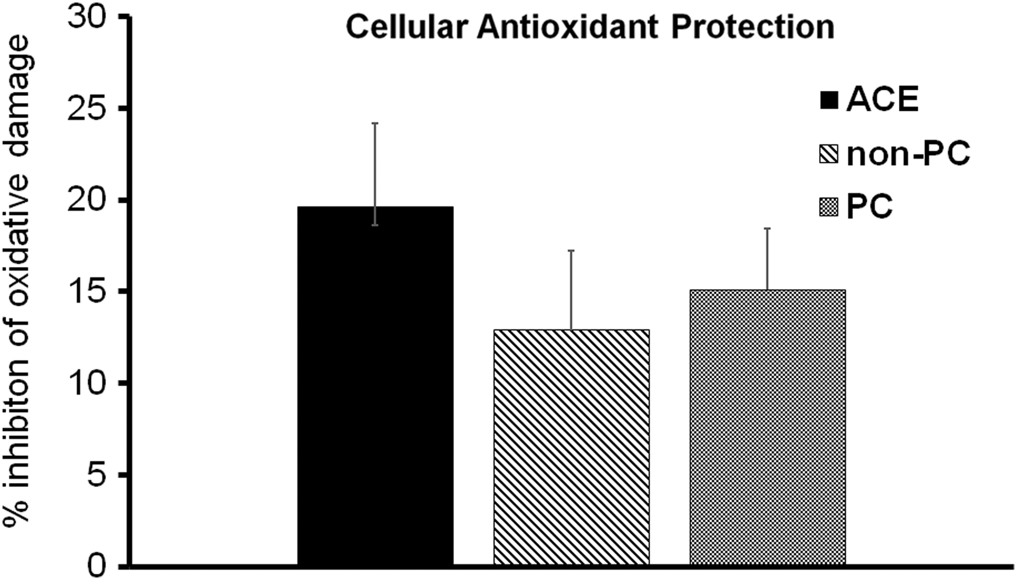
The cellular antioxidant protection (CAP) provided by the ACE, compared with PC and the non-PC fraction is shown for the dose of 4.2 mg/mL. Testing was performed using the CAP-e bioassay, where red blood cells are exposed to test products allowing the cells to absorb antioxidant compounds. After removal of unabsorbed compounds, the cells are then exposed to oxidative stress, and a reporter dye reflects the level of oxidative damage to the cells. The CAP provided by ACE was higher than the non-PC and the PC fractions separately, suggesting that the cellular protection from oxidative stress-induced damage had contribution from both PC and non-PC compounds. The difference between ACE and the two fractions did not reach statistical significance (p<.27).
The inflammatory PMN cell type was used in a bioassay to evaluate the free radical formation, specifically ROS. Both PC and the non-PC fraction contributed to reduced ROS formation under oxidative stress conditions, however, the ACE product containing both fractions performed best (Fig. 5). This specifically suggests that anti-inflammatory properties associated with ACE reflect contributing effects from both the PC and the non-PC fraction of ACE, and are not simply associated with PC alone.
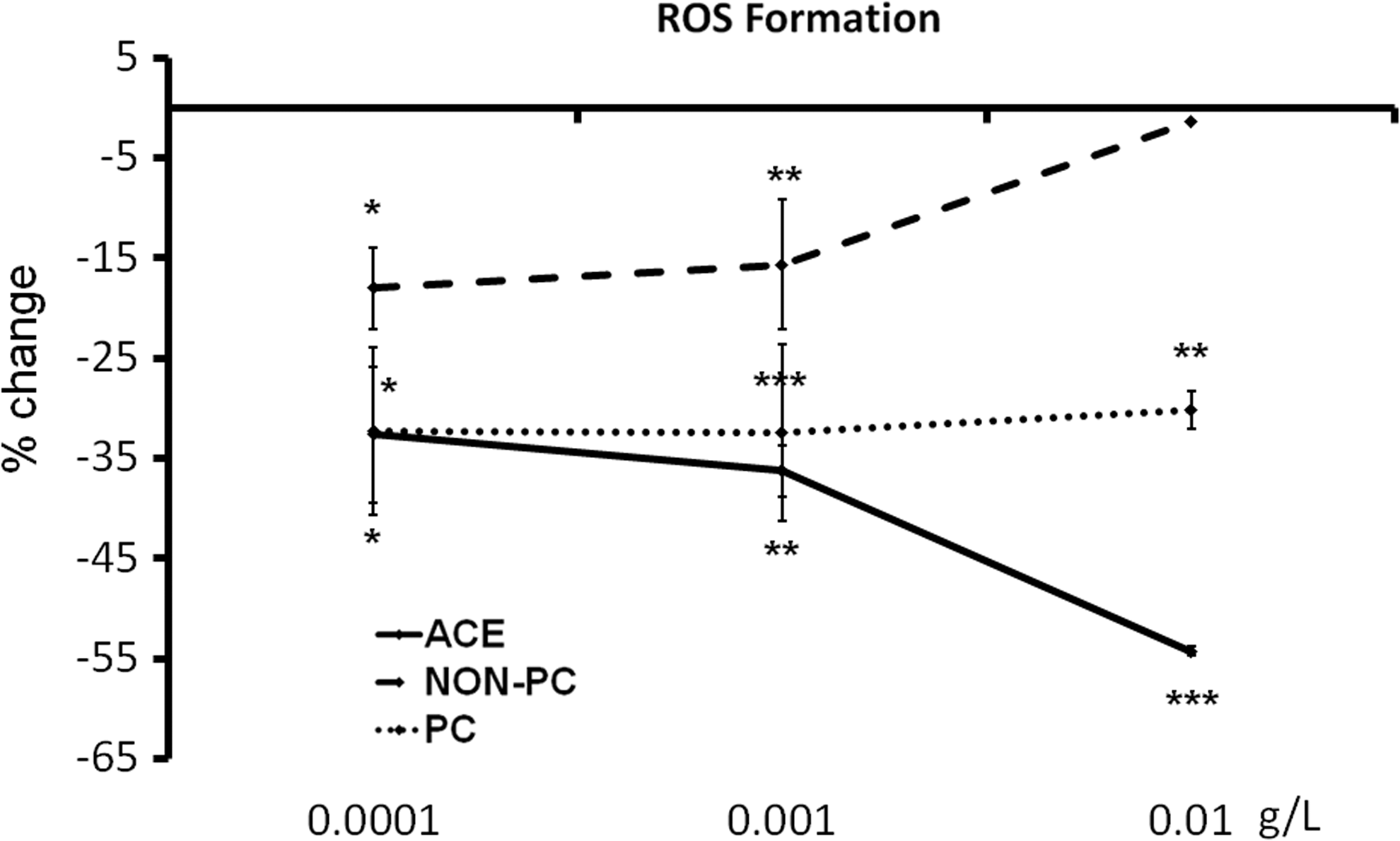
The reduction in inflammation-induced cellular production of reactive oxygen species (ROS) is shown for ACE, compared with PC and the non-PC fraction. Error bars reflect the standard deviation for each triplicate data set. The suppression of ROS formation was observed across a broad dose range for all three fractions. The cellular ROS production was mildly suppressed by the non-PC fraction, moderately by PC, and strongest by ACE. Levels of significance are indicated by asterisks: p<.05 indicated by *, p<.01 indicated by **, and very high level of significance if p<.001 is indicated by ***.
The effects of ACE, PC, and the non-PC fraction on clot formation and subsequent fibrinolysis was tested using the Euglobulin assay (Fig. 6). The addition of PC and ACE, and to a lesser degree the non-PC fraction, did not interfere with clot formation. In contrast, a stronger clot was formed, as seen by the higher maximum clot formation, happening immediately after the clot formation was initiated. Furthermore, despite a more robust clot formation in the presence of ACE, PC, and non-PC, the subsequent fibrinolysis, happening over the following hours, was resolved at the same time as untreated wells (Fig. 6A). A dose comparison showed that the more robust clot formation was statistically significant for PC and ACE at the two highest doses tested (Fig. 6B).
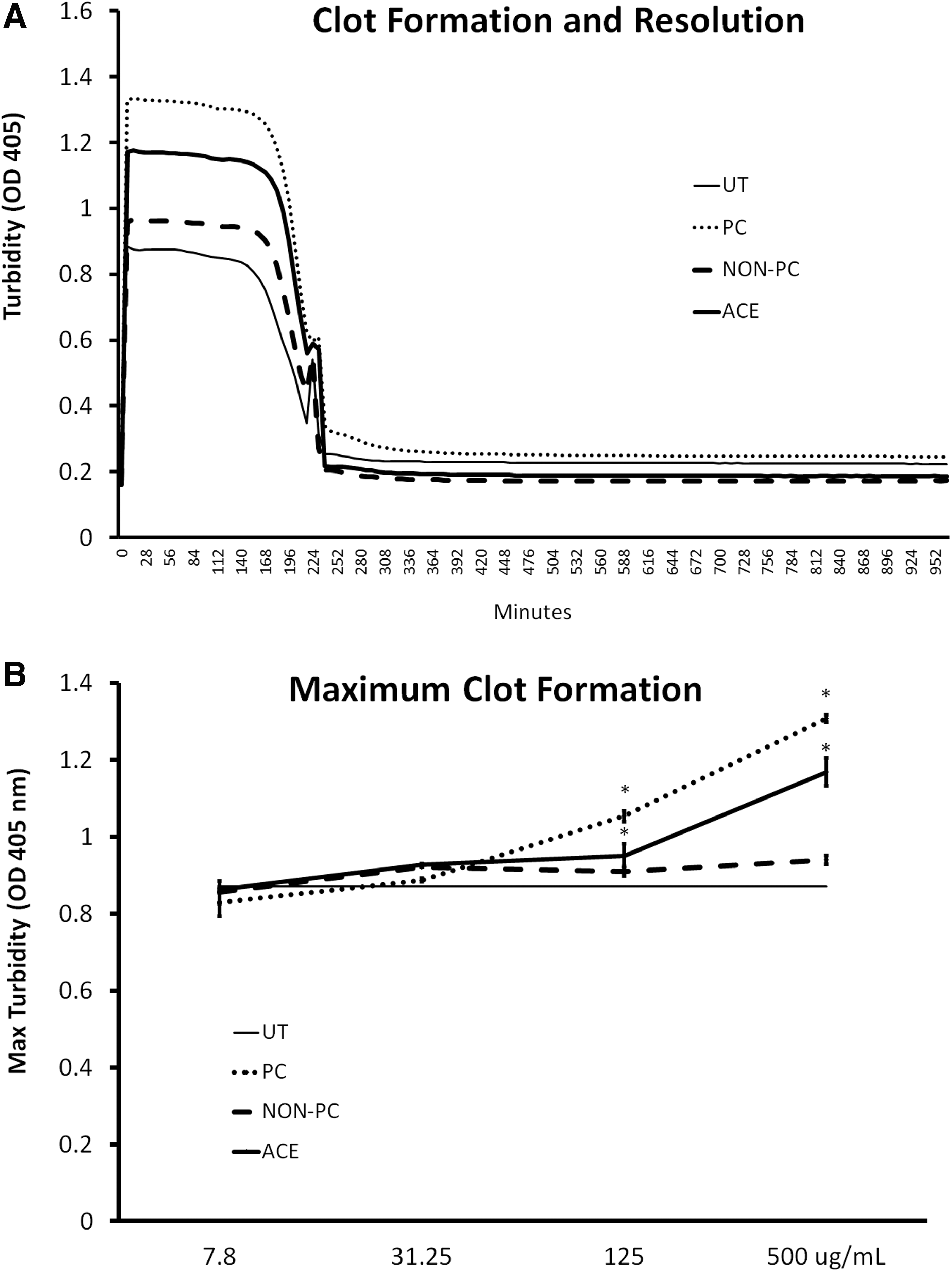
Clot formation and fibrinolysis in vitro.
Discussion
The health benefits of consuming edible cyanophyta has been documented in models of antioxidant status, 2 lipid health, 3 –5 and metabolic disorders. 6 Much of the biological activity has been suggested to be associated with the known COX-2 inhibitor PC. The data reported in this study are, to our knowledge, the first to specifically document complementary biological effects by non-PC compounds in the aqueous extract of Arthrospira. The presence of inhibitors of the Lipoxygenase enzyme in the non-PC fraction is interesting in light of the pharmaceutical efforts in pain management, where new approaches in the management of inflammatory conditions are being designed around simultaneous inhibition of both COX-2 and Lipoxygenase enzymes. 26,27 Consumption of ACE may, therefore, offer a nutraceutical method for the synergistic inhibition of these two enzymes.
In addition to the enzyme inhibiting properties, PC accounted for a major proportion of the antioxidant properties of ACE, with some contribution also from the non-PC fraction. Furthermore, beyond a simple antioxidant capacity, ACE inhibited the free radical formation by inflammatory PMN cells, thus further reducing oxidative stress.
Based on in vitro observations it has been previously suggested that PC may have value as a blood thinning agent, as PC can, under some circumstances, lead to reduced platelet–platelet aggregation, platelet activation, and platelet membrane fluidity. 28 However, we suggest that this interpretation may be too simplistic. Whereas it has been demonstrated in vitro we suggest that these effects must be interpreted as downstream effects of the initial anti-inflammatory effects, rather than as a separate effect that may potentially affect blood coagulation. Our data show that the presence of PC led to stronger clot formation in the euglobulin assay, whereas the subsequent natural fibrinolytic dissolution of the clot was unaffected. Further studies are warranted to get a better understanding of whether the in vitro data on blood clotting from our team and other researchers extend to the in vivo situation.
In conclusion, synergistic effects from compounds in the non-PC fraction of ACE increased the antioxidant and anti-inflammatory properties of ACE above the effect of PC alone. These aspects include the antioxidant capacity and the CAP, as well as the anti-inflammatory effect seen by the inhibition of ROS formation by inflammatory cells. Thus, the Arthrospira extract ACE holds promise as a nutritional support against chronic inflammatory conditions, 21 and the data suggest that purified PC may hold less promise than a more complex cyanophyta extract such as ACE. Clinical studies on ACE consumption are needed to expand on the in vitro observations, and examine the effects of ACE consumption in situations of pain, inflammation, and cardiovascular health.
Footnotes
Acknowledgments
The study was performed at NIS Labs, an independent contract research lab specializing in natural products research, located in Klamath Falls, OR, USA. The work was sponsored by Cerule LLC, Klamath Falls, OR, USA.
Author Disclosure Statement
G.S.J., K.F.B., and V.L.A. are employees of NIS Labs; none of these authors have any financial interest in the subject matter. J.G. and A.E. are employed by Cerule LLC, the sponsor of this study.