
Abstract
We describe a case of a 22-year-old male who presented to our facility 1 hour after a snake bite, which he identified as the desert black snake. He presented with severe weakness and respiratory distress. He was treated with polyvalent antivenom and observed in the Intensive Care Unit (ICU) with resolution of his respiratory symptoms. He developed paresthesias locally around his wound and later complained of diplopia. Two days later, he had total resolution of his symptoms. This is one of the only clinical reports of neurotoxic effects after Walterinnesia morgani envenomation.
Introduction
The Desert Black Snake (Figure) is a member of the Elapidae family. It is native to the sandy areas of the Middle East, including Kuwait, Saudi Arabia, Iran, Iraq, Jordan, Syria, Lebanon, and Egypt. There have been relatively few descriptions of human clinical effects of such snake bites, with most of the characterization of the venom from laboratory studies and in vitro experience. While the venom has been shown to have potent neurotoxic effects in laboratory studies, most clinical reports are limited to local reactions. 1
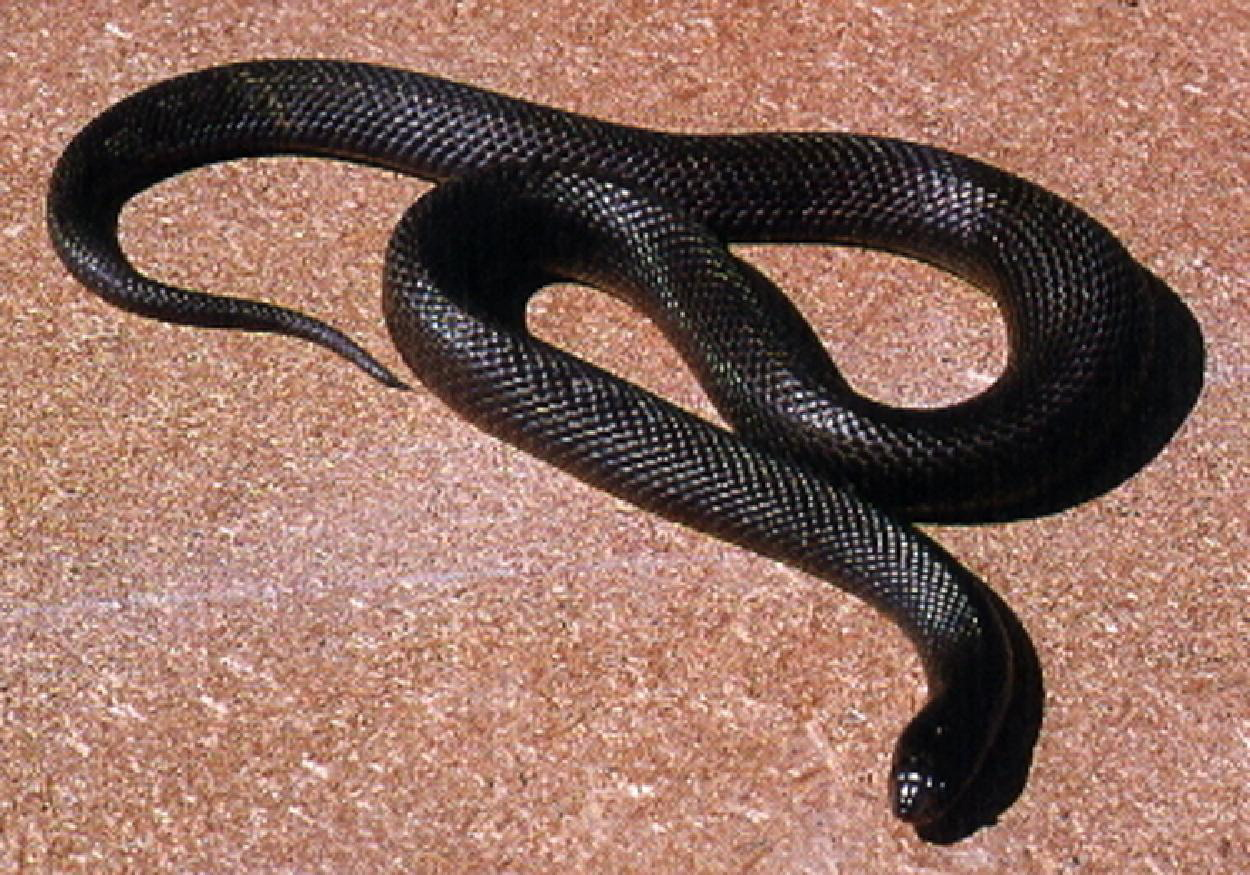
Black Desert Snake (photograph). Ahmed A. Egyptian Wildlife (internet). National Hepatology and Tropical Medicine Research Institute. Cairo, Egypt. c2005.
Case Report
A 22-year-old Iraqi male presented to a US Army facility in Baghdad with marked difficulty breathing 1 hour after a snake bite to his left lower leg. He was working along a construction wall outside of the military compound and happened upon the snake when it struck him. He was initially seen at a local clinic where an attempt was made at sucking the venom, and a tourniquet was applied above the wound. The patient began having respiratory difficulty and was given 1 vial of an unknown antivenom and 100 mg of hydrocortisone IV. He was immediately transported to the local US Army facility for further care. On presentation to the emergency department, he complained of moderate shortness of breath and weakness. On exam, his vital signs were normal except a moderate tachypnea. On arrival, he was treated with ipratropium bromide and albuterol sulfate nebulizer treatments without improvement. He was awake, but was in moderate respiratory distress. Formal cranial nerve testing was not performed before the administration of antivenom due to the patient's distress. His lungs were clear to auscultation, although with rapid, shallow breaths. His heart had a regular rate and rhythm. He exhibited global decrease in strength, with his legs (2/5) affected much more than his arms (4/5). His left medial calf had a 5 cm area of erythema, mild induration, and tenderness around 2 small puncture wounds. When questioned, he described it as a large black snake and was shown an instructional poster containing pictures of the local venomous reptile population. 2 He clearly identified the Desert Black Snake as the responsible reptile. He had a normal electrocardiogram, a mild leukocytosis to 14 000 white blood cells/μL, and normal prothrombin time and partial thromboplastin time. He was treated with 3 additional vials (30 mL) of antivenom (polyvalent equine snake antivenom, The Antivenom & Vaccine Production Center, Riyadh, Saudi Arabia) with gradual improvement in his respiratory distress over the next hour. He had no evidence of an adverse reaction to the antivenom. While his respiratory status improved, his motor strength remained diminished. He was admitted to the ICU for further observation and frequent neurologic reevaluation. Twelve hours later, the patient was clinically improving with no respiratory distress and a return of normal motor strength to all extremities. An area of numbness around the bite on his left calf remained. Twenty-four hours after the envenomation, the patient complained of diplopia. Evaluation by the facility optometrist found that his visual acuity and his alignment were normal. Over the course of the next 24 hours, his diplopia improved and he had no further complaints. His mild leukocytosis resolved after 48 hours. The erythema and induration of his left calf wound resolved. He had no evidence of renal, hepatic, or metabolic disturbances during his hospital stay. He had no adverse reaction to the antivenom during his hospital course. He was discharged on hospital day 3 in excellent condition.
Discussion
The Desert Black Snake, belonging to the Elapidae family, is indigenous to the sandy areas of the Middle East, including Kuwait, Saudi Arabia, Iran, Iraq, Jordan, Syria, Lebanon, and Egypt. 3 It is also the only Elapid found in Israel. It may attain a length of 120 cm and is glossy black in color, sometimes with a shade of brown. 4 The Desert Black Snake has recently been subdivided into 2 species, Walterinnesia morgani, of the eastern areas of Iran, Iraq, and eastern Saudi Arabia, and Walterinnesia aegyptia, of the western areas of Egypt, Israel, and western Saudi Arabia, based on several morphological distinctions. 5 One of the most significant of the distinctions is the juvenile color pattern of Walternnesia morgani, which displays red crossbars on a black body, while W aegyptia remains solid black in its juvenile form.
Elapidae venoms usually possess neurologic and cardiac toxicity, which can progress to respiratory arrest, arrhythmias, and death. 6 Less commonly, Elapid venoms may also lead to local tissue injury and hematologic abnormalities. Viperidae venoms typically produce local tissue destruction and hematologic abnormalities, but they may also have cardiotoxic and neurotoxic effects. Viperidae snakes, however, rarely cause respiratory arrest. Elapid bites are characterized by local and systemic poisoning, with local reactions mainly limited to pain and swelling at the site and, later, variable degrees of necrosis. These are typically less severe than those of the pit vipers (Crotalinae).
While clinical cases of Desert Black Snake bites are fairly rare, the snake's venom has been studied and shown to be potently neurotoxic and also possesses hemorrhagic activity. 4 In mice, at doses 3 times the “Lethal Dose, 50%” (LD50), the mice would experience increasing muscle weakness, impairment in respiration, convulsions, paralysis of hind legs, and death by 60 minutes. At higher doses, the toxic effects appeared more rapidly. The venom of Walterinnesia is known to contain 3 neurotoxins (W-III, W-IV, and W-V), which act on the postsynaptic receptors to block neuromuscular transmission. 7 These are curaremimetic neurotoxins with a high affinity to the acetylcholine receptor. While one of the neurotoxins (W-III) is reversible with a lower affinity for the acetylcholine receptor, the other 2 (W-IV and W-V) have been shown to be irreversible and far more potent. In envenomated mice, clotting time remained normal and no fibrinolytic activity was observed, but there did appear to be evidence of varying degrees of congestion and hemorrhage in the liver, kidneys, brain, and lungs. 4 Gitter et al has shown, in vitro, Walterinnesia venom possesses protease, phosphatidase A, hyaluronidase, 1-amino acid oxidase activity, weak anticoagulant, and some fibrinolytic activity.
While detailed clinical descriptions of neurotoxic effects of Desert Black Snake envenomations are not available, there have been descriptions of other elapid envenomations with neurotoxic effects. Sweating, numbness, paresthesias, convulsions, coma, muscle fasciculation, muscle weakness, and respiratory arrest are among the clinical sequelae. 6 Respiratory arrest is the primary cause of death in elapid bites, and this may occur within 15 to 30 minutes, or may be delayed up to several hours.
Most published cases of bites of the Desert Black Snake involve more local reactions. In one case report of envenomation, a 22-year-old female was bitten on the finger of her left hand while she was sleeping outside during the night. 1 The patient exhibited swelling of her left hand to her wrist with pain extending up her left arm. She did not exhibit any systemic effects except mild agitation. She had a leukocytosis with a normal coagulation panel. An EKG on admission showed sinus tachycardia and premature ventricular contractions, which normalized after 4 hours. Other reports have shown similar benign courses, including a case series of 4 reported bites in Israel, resulting in localized pain and swelling, fever, generalized weakness, nausea, and vomiting. 8 A more severe reaction was noted by Corkill, who described a snake charmer of the Iraq Army that tried to capture a black snake. 9 He was bitten and died 6 to 10 hours later. Due to his description of the snake to his companions and the fatal outcome, the snake was thought to be a Hoodless Cobra or Desert Black Snake. The differences between the known neurotoxic effect and these more benign cases might be explained by differences in the venom of W aegyptia and W morgani and their respective geographic habitats.
The relatively benign clinical courses of these bites have been attributed to both the non-aggressive behavior of the snake as well as the mechanism of the bite. 1 While other snakes will quickly strike the victim, envenomating them in a relatively small period of time, the Elapidae family has a slower envenomation process, requiring an extended period of contact between the snake and the victim to deliver the adequate quantity of toxin. The volume of available venom can be affected by recent feedings as well. 6 While 30% of crotalid bites result in no envenomation, 50% of elapid bites will be dry. 6
While the identification of the snake by patient report without physical confirmation remains inconclusive, the authors are confident of the identity of the snake due to the absence of other similar-appearing snakes in the region of central Iraq and the rapid improvement in symptoms with antivenom. The presence of 2 identical marks identified on the leg suggests a snake bite instead of another possible source of venom. Of the venomous snakes native to Southwest Asia, the Central Asian Cobra—or Naja oxiana—is the only other large dark-colored venomous snake, and it is not endemic to Iraq. 2 The remainder of the venomous species found in Iraq are viperid without known neurotoxic venom, and they are not similar in appearance. Dolichophis jugularis, or the Whip Snake, is a similar-appearing, aggressive, nonvenomous snake that is known as “urbid” or “abrid” locally, and is frequently confused for the Desert Black Snake.9,10 The Whip Snake can be found in northeastern Iraq but is not endemic to central Iraq where this envenomation occurred. 11
Anaphylactic shock remains a remote possibility. The incidence of anaphylaxis in envenomation is believed to be only 1%, making it less common than other physiologic effects. 6 While there is a possibility that the reaction in this case was secondary to anaphylaxis, the absence of wheezing, urticaria, laryngeal edema, and hypotension make anaphylaxis unlikely. In addition, the patient's rapid improvement with antivenom alone, without the use of epinephrine and a histamine blockade, suggests a neurotoxic effect instead of an anaphylactic event.
The antivenom used in this case was a polyvalent equine antivenom from Saudi Arabia, consisting of a highly purified preparation of F(ab')2 fractions of immunoglobulins against 6 venomous Saudi snakes. 12 The Saudi polyvalent antivenom is specific to neutralizing the toxins of Bitis arietans, Cerastes cerastes, Echis carinatus, Echis coloratus, Naja haje, and W aegyptia. In addition, it has shown to have a wide spectrum of activity against other Middle East and North African snakes, including Bitis caudalis, Bitis gabonica, Naja melanoleuca, Naja naja, and Naja nigricollis. This antivenom is indicated if signs of systemic toxicity are present after a Middle East or North African snake bite. It should be given as soon as possible and it is generally given as 40 mL (four 10 mL vials) diluted in 5 mL of physiologic isotonic fluid/kg body weight. This is infused slowly over a period of 30 to 60 minutes. Alternatively, it can be given IV, undiluted, at a rate of 4 mL/min. There is no difference in the incidence or severity of antivenom reactions with either method. 12 In addition, since the dosing of antivenom is based on the amount of antivenom in the body and not the volume of distribution, the dosing is not based on weight, and children should receive the same dose as adults and may require a larger dose. The antivenom dose can be repeated every 4 to 6 hours as needed for clinical symptoms. Potential reactions should be identified after any antivenom administration, including anaphylaxis in 5% to 25% of cases. 6 These Type I hypersensitivity reactions are typical within the first 10 to 180 minutes of administration. Later consequences include Type III hypersensitivity, or “serum sickness,” occurring 5 to 24 days after antivenom administration, with a variable incidence depending on the antivenom. Typically, horse serum-based products tend to be more immunogenic than sheep-based antivenom. 6 In addition, Fab immunoglobulin fragment antivenoms have a lower rate of allergic reactions than IgG or F(ab')2 fragment antivenom.
This is one of a few descriptions known to the authors of a clinical case of the neurotoxic effects of the Desert Black Snake. Previous case reports, limited to local effects without systemic toxicity in the majority of cases, are not consistent with the known toxicology of the Walterinnesia venom. In our case, the local effects were fairly mild, but the neurologic symptoms were severe, more representative of what is known about this neurotoxic venom. In addition, this patient had a significant leukocytosis which cannot be explained by the Walterinnesia venom, but may be the result of demargination of white blood cells from a sympathetic effect after the envenomation. It remains unclear why there are such varied clinical courses, but this is likely due to both the nonaggressive nature of the snake and the prolonged contact required for envenomation, resulting in variable amounts of venom injected with bites. In addition, it is possible that there is some variation in venom toxicity as well as the known morphological variations between W aegyptia and W morgani, although this has not been specifically studied.