
Abstract
Objective/Background
Tactile perception is a basic way to obtain and evaluate information about an object. The purpose of this study was to examine the effects of tactile perception on brain activation using two different tactile explorations, passive and active touches, in individuals with chronic hemiparetic stroke.
Methods
Twenty patients who were diagnosed with stroke (8 right brain damaged, 12 left brain damaged) participated in this study. The tactile perception was conducted using passive and active explorations in a sitting position. To determine the neurological changes in the brain, this study measured the brain waves of the participants using electroencephalography (EEG).
Results
The relative power of the sensory motor rhythm on the right prefrontal lobe and right parietal lobe was significantly greater during the active tactile exploration compared to the relative power during the passive exploration in the left damaged hemisphere. Most of the measured brain areas showed nonsignificantly higher relative power of the sensory motor rhythm during the active tactile exploration, regardless of which hemisphere was damaged.
Conclusion
The results of this study provided a neurophysiological evidence on tactile perception in individuals with chronic stroke. Occupational therapists should consider an active tactile exploration as a useful modality on occupational performance in rehabilitation training.
Introduction
Stroke is due to a cerebral vascular accident that can induce motor and sensory dysfunctions in individuals (
Tactile perception is one sense composing touch, a complex system, with pain perception, temperature perception, proprioception, and kinaesthetic perception (
Neurophysiological research is in progress to evaluate the effectiveness of tactile perception, including the addition of active movement (
As mentioned, previous studies have examined the dynamic effects of the types of tactile exploration on brain activation and movement learning in healthy children and the adult population (
This study examined the effects of tactile perception on brain activation using electroencephalography (EEG) on two different tactile exploration methods—passive and active touches, in individuals with chronic hemiparetic stroke, and compared the differences of brain responses between those with right and left brain damages. We hypothesized that there would be a significant difference in the two different tactile perception on brain activation during movement, and that the brain activation of the impaired side of the brain (e.g., right or left) would be affected by the two different tactile explorations during movement in the chronic hemiparetic stroke population.
Methods
Participants
Twenty people who were diagnosed with stroke (8 right brain, 12 left brain) participated in this study. The inclusion criteria included: (a) adults who have received a diagnosis of hemiparetic stroke; (b) the onset was > 1 year previously; (c) adults who received 23 points or more on the Mini-mental State Examination (MMSE) (
Procedure
To minimize any external bias during the experimental process, this study requested that the participants maintained their sitting position without moving and with closed eyes until the experiment was finished. This study then measured their brain waves. The tactile exploration was conducted while the participants were in a sitting position. For the first 3 minutes, we measured the participants' resting EEG, and applied the tactile exploration. The tactile exploration was conducted using two methods: passive exploration and active exploration. The passive exploration was conducted while a tactile board was moved by a therapist and felt by the participant. The active exploration was conducted while the participant actively moved a tactile board (
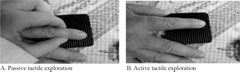
(A, B) The experimental procedure of two different tactile explorations.
Outcome measure (experimental equipment)
To determine the neurological changes in the brain, this study measured the brain waves of the participants using a CANS 3000 QEEG-8 (Laxtha, Inc., Daejeon, Republic of Korea), which measured the participants' sensory motor rhythm (SMR) to compare the effects of the tactile exploration tasks. The SMR is activated by somatosensory stimuli and active movement. In addition, alpha, mu, and median (high) beta rhythms were corrected with consciousness, attention without movement, and cognition respectively. This study aimed at evaluating the SMR because it was related to attention ability in the frontal and occipital lobes, and that the alpha and median beta rhythms might not show relationship with the active movement relatively compared with SMR and mu rhythm. EEG was recorded using Ag/AgCl cup electrodes attached to the scalp at eight active sites (Fp1, Fp2, C3, C4, T3, T4, P3, P4), according to the international 10/20 system. In addition, a relative power analysis was conducted on the values at the eight active positions.
Data analysis
For the EEG analysis, this study collected data on the relative power of each participant SMR. A comparison of the relative power spectrum calculations was conducted using the research task conditions and the Fast Fourier Transform. The SMR was determined using the A1 and A2 sites as the standard electrodes. For a statistical analysis, this study conducted a descriptive analysis and Wilcoxon's signed-rank test using PASW version 18.0 (SPSS Inc., Chicago, IL, USA). The significance was set at p < .05.
Results
Table 1 shows the characteristics of the participants. In the right brain damaged stroke group, average age was 64.9 ± 2.9 years, average score of MMSE was 26.8 ± 2.3 points, and average score of Modified Barthel Index was 69.8 ± 29.9 points. In left brain damaged stroke group, average age was 66.4 ± 3.3 years, average score of MMSE was 25.0 ± 2.2 points, and average score of Modified Barthel Index was 69.0 ± 26.5 points (Table 1).
General Characteristics of the Participants (n = 20).
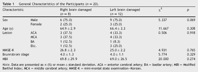
Note. Data are presented as n (%) or mean ± standard deviation. ACA = anterior cerebral artery; BA = basilar artery; MBI = Modified Barthel Index; MCA = middle cerebral artery; MMSE-K = mini-mental state examination—Korean.
This study measured the relative power of the SMR on prefrontal, central, temporal, and parietal lobes in both the right and left hemispheres to compare the effects of a tactile exploration task using passive and active touches. In participants with right hemispheric damage, the active tactile exploration induced higher relative power of the SMR on the central, temporal, and parietal areas in the right hemisphere compared to the relative power in the passive tactile exploration task. The relative power of the SMR was greater only in the central area during the active tactile exploration compared to the relative power during the passive tactile exploration; however, no brain areas were significantly different between the active and passive tactile explorations (Table 2).
Value of Sensory Motor Rhythm during Passive and Active Explorations of Participants (n = 20).
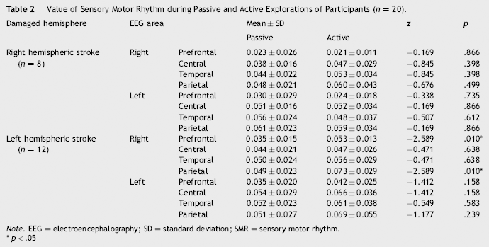
Note. EEG = electroencephalography; SD = standard deviation; SMR = sensory motor rhythm.
p < .05
In those participants with left hemispheric damage, the active tactile exploration induced a greater relative power of the SMR on all four sites, including the prefrontal, central, temporal, and parietal areas in the right hemisphere, compared to the relative power in the passive tactile exploration condition. However, the relative power of the SMR was significantly higher on the prefrontal and parietal areas in the right hemisphere. Although the active tactile exploration showed a greater relative power of the SMR in the left hemisphere compared to the relative power in the passive tactile exploration condition, none of the sites were significantly different between the two tactile exploration tasks (Table 2).
Discussion
This study examined the effects of two different tactile explorations, passive and active touches, on EEG brain activation in individuals with right and left brain damage due to stroke. The results of this experiment revealed two significant findings. First, the relative power of the SMR on the right prefrontal lobe was significantly greater during the active tactile exploration compared to the relative power during the passive exploration in the left damaged hemisphere. Second, the relative power of the SMR on the right parietal lobe was significantly greater during the active tactile exploration compared to the relative power of the passive exploration in the left damaged hemisphere. Most of the measured brain areas showed nonsignificantly higher relative power of the SMR during the active tactile exploration, regardless of which hemisphere was damaged.
Tactile exploration provides important sensory information about objects in ADLs because specialized sensory touch receptors perceive information about pressure, vibration, and movement during daily functions (
In recent studies, brain activations that depended on tactile recognition type, attention, and textures differed in the healthy adult population (
Previous studies have reported that prefrontal and parietal areas show a sustained response and greater activation during the active exploration of textures in healthy adults (
This study had a limitation in that it did not measure any subjective or behavioural responses after each trial of tactile exploration, so there may be a high variability in attention among the participants.
Conclusion
This study confirmed that the brain activation between passive and active tactile exploration is different in stroke patients with left, but not right damaged hemispheres. The SMR had a meaningful difference in this study because of its relationship to the active movement of the human body. This study confirmed that an activation of the brain waves is formed in the right parietal and frontal lobes in left damaged hemisphere. Those areas play key roles in the occupational performance and cognition related to information processing in humans. In future study, the therapeutic approaches using tactile exploration should incorporate brain measurements such as EEG to provide more evidence on the role of tactile perception in daily activities in training by occupational therapists.