
Abstract
Cytokine flow cytometry (CFC) is a multiparameter assay of antigen-specific T cell function, potentially useful in the monitoring of experimental vaccines and progression of infectious diseases and cancer. Automation of CFC assays would greatly facilitate their use in clinical trials and involves several components. We describe here the migration of these assays to 96-well plates, the use of sample-handling robotics, and the use of lyophilized antigen and antibody plates to help automate CFC. Together, these elements can produce an integrated system capable of walkaway automation of an entire assay, resulting in the reproducible processing of potentially hundreds of samples per day. Implementation of such systems has begun to be undertaken by our group and others.
Keywords
Introduction
Introduction to CFC Assays (Rationale)
Multiple methods have been developed to track antigen-specific T cells during immune responses. Some assays detect T cells based on the specificity of their T cell receptor (e.g., MHC-peptide tetramer 1 or dimer 2 binding). These assays are simple and powerful but require knowledge of both the MHC type of the donor and the immunodominant peptide epitopes comprising the response. Other assays detect T cells by their function (e.g., cytokine production or proliferation). These assays include enzyme-linked immunospot (ELISPOT) 3 and cytokine flow cytometry (CFC) 4 for cytokine production, and bromodeoxyuridine (BrdU) incorporation 5 and 5-(6-)carboxyfluorescein diacetate succinimidyl ester (CFSE) dilution 6 for proliferation. These assays are able to quantitate responses to complex antigens without regard to MHC type, although each assay can quantitate only T cells of a given function. Since the complexity and requirements of all these assays are different, not all of them are equally feasible for use in large clinical trials, where reproducibility, hands-on time, and ability to automate are prime considerations.
The most commonly used assays of antigen-specific T cells in clinical trials today are ELISPOT and CFC. This is likely a result of the fact that they are short-term assays (as opposed to BrdU incorporation or CFSE dilution), and they can detect immune responses to complex antigens without regard to donor MHC type (as opposed to MHC-peptide tetramer or dimer binding). Of the two assays (ELISPOT and CFC), CFC has the advantage of a highly multiparametric readout (flow cytometry), which allows multiple phenotypic measurements to be made on the cells in addition to measuring cytokine production. However, ELISPOT is considered a higher throughput platform because it is normally done in a 96-well plate format and can be read on an automated reader. Recent technical developments in CFC, however, have begun to bring similar advantages to this assay. 7,8 In fact, the case is made in this review that virtually complete automation of CFC assays is possible, making CFC an attractive assay for gathering high-content information on T cell responses in a high-throughput manner. Since the precise parameters that correlate with protection are largely unknown for diseases like HIV (for a review, see Maecker and Maino, 2003 9 ), the argument for collecting high-content information in clinical trials is a strong one.
Introduction to CFC Assays (Technical)
It is possible to capture secreted cytokines on the surface of the cells producing them 10 or for IFNγ and IL-10 to detect natural, low-level expression of cytokines on the cell surface. 11 However, the most common variation of CFC measures the intracellular accumulation of cytokine(s) in cells whose secretory pathway has been blocked by treatment with brefeldin A4 or monensin. 12 This latter method, often referred to as intracellular cytokine staining (ICS), has been applied to detection of antigen-specific T cells in both PBMC 4 and whole blood. 13
Activation of cells for CFC is deliberately short, typically 6 h, in order to minimize changes in the apparent frequencies of antigen-specific T cells arising from apoptosis or proliferation in longer term cultures. This activation can be done entirely in the presence of brefeldin A or monensin (for peptides and mitogens), or the secretion inhibitor can be added after an initial period of about 2 h (for whole protein antigens), to allow for antigen processing and presentation. Activation can be carried out in an incubator or water bath, with or without CO2 supplementation. The activation can be halted by decreasing the temperature to 4 or 18 °C14 (18 °C is recommended for whole blood to prevent platelet activation and resultant cell clumping). By using a programmable water bath or incubator, activation can be achieved without requiring the presence of the operator at the end of the desired incubation time.
Processing of activated cells for CFC usually begins with a brief treatment with EDTA to dislodge cells that have adhered to the vessel wall. Thereafter, cells are treated with fixative and detergent to prepare them for intracellular staining. Antibodies to some cell-surface antigens must be added prior to fixation, while others can be used together with cytokine-specific antibodies in a single staining step. Finally, it is possible to freeze cells at –80 °C immediately after fixation in order to batch samples for later analysis. 14
Acquisition of samples on the flow cytometer should generally be done within 24 h of staining. Various gating methods are in use by different groups, and automated analysis can be performed with some software packages. 7 This involves the use of cluster-finding algorithms that are built into the analysis template and allow regions to be drawn in a data-dependent manner. Statistical results of each sample's analysis can also be downloaded directly into a spreadsheet file, which avoids the added time and potential errors associated with manual data entry into a spreadsheet or database.
Regardless of the gating strategy used, interpretation of CFC results depends on the number of events collected and the difference between the negative control stimulation and the test stimulation. A power calculation can be done to determine whether a sufficient number of events were collected for this difference to be statistically significant. 15
CFC Assays in Plates
The original publications on human antigen-specific CFC staining used protocols based on stimulation in 15-mL conical tubes and processing in 5-mL round-bottom tubes (compatible with most flow cytometers). 4,13 However, the authors and others have adapted both the stimulation and processing of CFC assays to 96-well plates (Fig. 1), 8 and we recently published a comparison of plate- and tube-based methodologies. 7 Results were comparable between plate- and tube-based methods for both whole blood and PBMC.
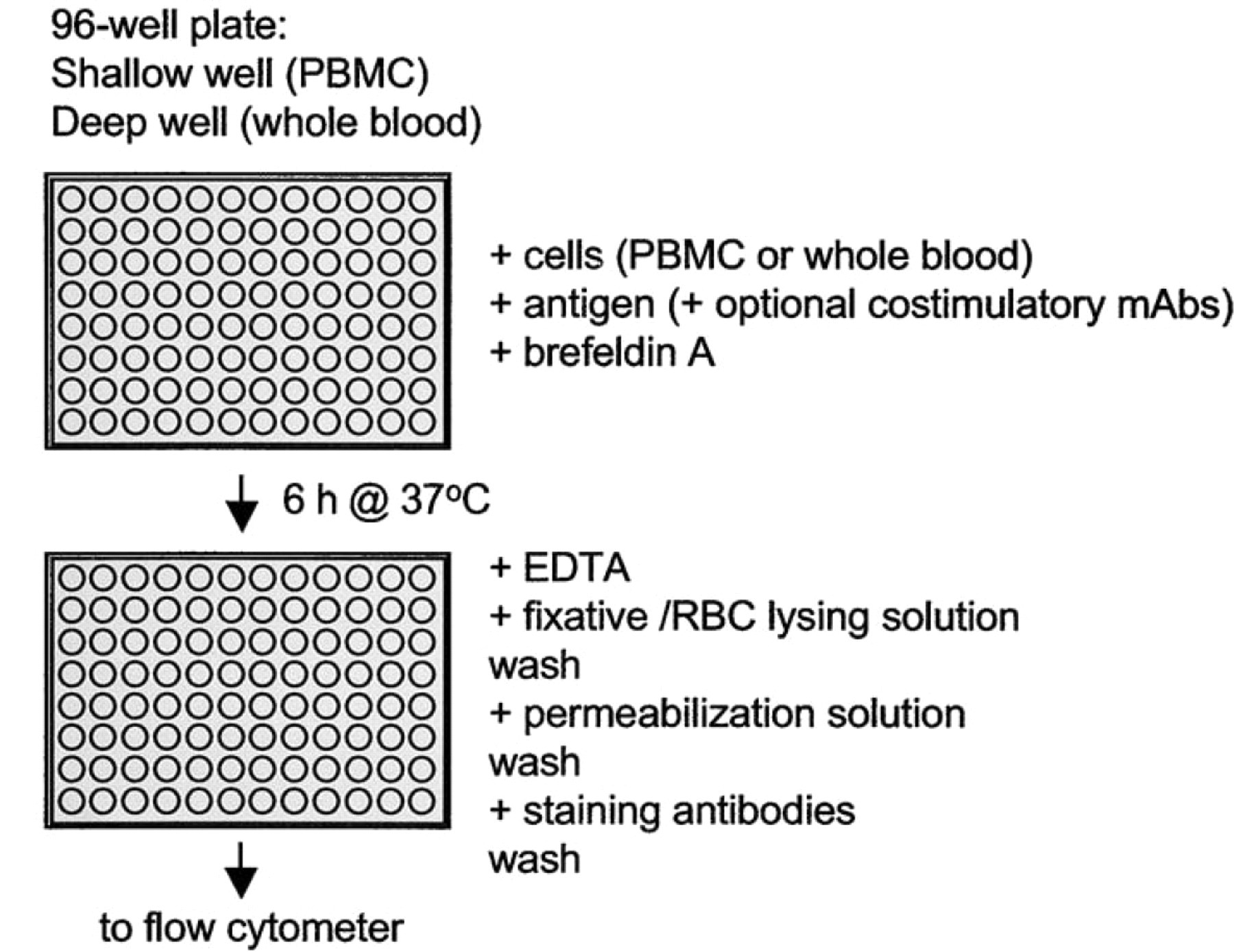
Schematic of a plate-based CFC protocol. The same plate can be used for activation, processing, and data acquisition; parallel processing of 96 samples saves considerable operator time compared to tube-based protocols. The authors have stimulated 0.5–2×10⁶ PBMC per well with equivalent results, or 0.2-mL whole blood (in 96-well deep-well plates). Up to 1 mL of whole blood, or higher numbers of PBMC, can be stimulated in 24-well deep-well plates.
Plate-based CFC assays provide multiple advantages to throughput and reproducibility. By using conical or round-bottom plates, the same vessel can be used for activation, processing, and acquisition of samples (provided the flow cytometer is equipped with a plate loader). This method avoids cell loss associated with transfer from one vessel to another and reduces processing time. Also, the advantages of plate-based sample handling (parallel centrifugation of all samples, multichannel addition of reagents, and aspiration of supernatants) mean that 96 samples can be processed in about the same time as one or a few tubes. This should result in increased reproducibility across large sample sets that otherwise would suffer from differential incubation times if processed serially in tubes. Finally, the rigid matrix configuration of a plate allows more intuitive experimental design with reduced probability for sample-switching errors.
Robotics for Plate-Based CFC
The migration of CFC assays from tubes to plates allows investigators to capitalize on the growing family of instruments, workstations, and larger automated systems developed for cell-based assays in the drug discovery environment. Unlike drug discovery where speed is extremely important, the primary goal of these automated systems is robust performance of a complicated assay without manual intervention. We need to handle tens or hundreds of samples, not thousands, and we need systems that generate data on humans, not compounds, so that reliability supercedes throughput as a priority.
The authors and others have assembled such systems, using components from various manufacturers, with priorities on robust performance of various aspects of the overall sample handling process 16,17 (and Barry Bredt, personal communication). One such system is depicted in Figure 2A. It includes a robotic arm that can move plates between a pipetting station, a heat block/incubator, an indexing plate centrifuge, and a plate-loading cytometer. With properly designed protocols, this system can execute all of the steps of a CFC assay, from aliquoting whole blood or PBMC into plates for activation to acquisition and analysis of flow cytometry data generated from those plates. Results are well correlated with manual CFC results 16 (see also Fig. 2B), and it is expected that this correlation will improve further with additional protocol refinements.
Common requirements of any system for automated CFC are likely to include centrifugation of cell suspensions to allow serial incubations and washes; fluid handling with ∼10% precision in the tens to hundreds of microliter range; 37 °C incubation for activation; and plate robotics to move plates from various subsystems, including a centrifuge, pipettor, cytometer and incubator(s). Commonly desired subsystems may also include a barcode reader, cap piercing for the initial distribution of samples from blood draw tubes to plates, pick-and-place fluid handlers and manipulators to handle reagent distribution, chilling to 18 or 4 °C to stop activation and assay reagent degradation, biohazard containment, and light minimization to protect photosensitive reagents. As an alternative to centrifugation, filter bottom plates may be used with vacuum aspiration. 17 Our experience suggests that these can result in a high level of cell loss if the plates are not kept continuously wet during aspiration, a process that is difficult to precisely control.
A summary of experiences with these systems is probably similar to other such automation enterprises. Precise protocol definition is key to success, and every modification is painful. Scientists who are accustomed to easily modifying assays will be frustrated by time-consuming reoptimization of protocols and the surprising constraints in system hardware and software. For example, in processing one set of samples for CFC analysis, there are 14 different pipetting subprotocols that are called serially with centrifugation steps in between. A change in the amount of fluid added to the plate, either as a change in the number of filled wells or in the amount of fluid per well, changes requirements on the weight of a balance plate for that centrifugation. In some cases, the simplest way to handle that occurrence is to maintain constant spin weights, which means that a change in any step requires a change in all steps. In other cases, step-specific changes are required, which trigger management of a second or third balance plate, adding further complication. Walkaway automation of such a complicated assay is a high standard and requires a real investment in development time and expertise. When successful, however, such systems can generate data, which is not only correlated with manual sample handling (Fig. 2B), but should also be subject to increased precision due to better reproducibility of automated vs. manual procedures.
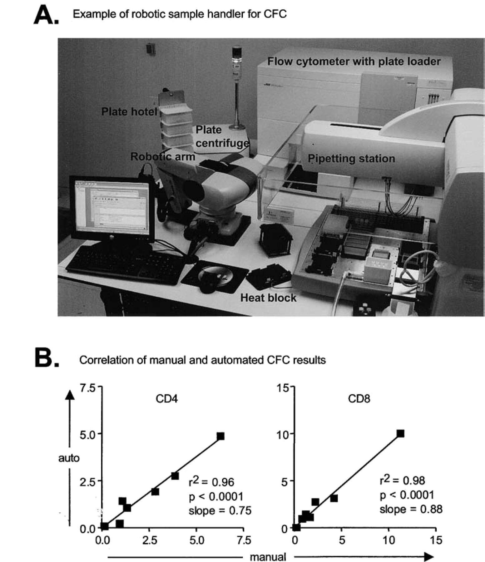
(A) Example of a robotic workstation for automated activation, processing, and acquisition of CFC samples in plates. Major components are as labeled. Note that the heat block pictured was determined to be less effective than a conventional incubator for sample activation. (B) Correlation of the percentage of CD69 + IFNγ+ cells obtained after activation and processing done manually vs. on the automated workstation shown in (A). Results are for both SEB and CMV peptide mix activation of whole blood from four donors (unstimulated background subtracted).
Lyophilized Reagents in Plates
While the power of plate-based flow cytometry is the generation of a rich array of measurements comparing many patients with many stimuli and many response features, implementation of such powerful strategies can be daunting. One aspect of CFC automation that is particularly challenging is the control of the experimental environment and dispensation of some of the reagents. Fluorochrome-conjugated monoclonal antibodies and specific antigen peptide cocktails are expensive and, at least in principle, can be sensitive to temperature, light, and cross contamination. Common experimental paradigms will require dense patterns of distribution of these reagents within single plates, with either many different antibody or antigen cocktails, or both, per plate. When added to complicated distribution patterns of individual patient blood samples and many patient samples per plate, the pipetting requirements of an automated experiment certainly become rate limiting and even conceptually limiting at the experimental design phase.
To simplify those aspects of experimental design that are separate from the blood samples, the authors have migrated our assays to preformatted, freeze-dried antigen and antibody plates. Work to date suggests that performance of antibody-fluorochrome conjugates commonly used in flow cytometry can be reasonably maintained in optimized freeze-drying protocols, and the shelf life of well-sealed preparations can be measured in years. Antigen cocktails are less well studied to date, but early indications are also encouraging (Fig. 3). For protein antigens, lyophilization protocols similar to those used for antibody cocktails seem appropriate. Antigen peptide cocktails, including those with Brefeldin A, are unusual in that they will typically require DMSO at some concentration to maintain solubility. Solvents require modification of the freeze-drying procedure in order to achieve classic drying without the collapse of the product, but in functional studies, even collapsed product still seems to maintain good activity.

Comparison of CFC results using liquid vs. lyophilized reagents in plates. An example of a response to CMV pp65 peptide mix activation is shown, gated on CD3+CD4- cells (A) or CD3+CD4+ cells (B). Differences in cytokine staining intensity seen in this example are not typical of all experiments done with lyophilized vs. liquid antibodies.
Preconfigured plates of freeze-dried antigen and antibody allow for complex assay configurations to be reliably repeated many times over the course of a study with minimal effort. For automated CFC assays, they greatly simplify pipetting protocols and save sample-processing time, while reducing concerns about reagent degradation on the robot. In addition, lyophilized reagents offer real advantages for long-term studies where stability of reagents is important. Particularly in clinical trials, reagent and laboratory variables can obscure subtle but important patient data trends over the several years of most studies.
Conclusion
Automation of CFC assays is reasonable given currently available technologies and devices. Components of the automation process include the migration of CFC assays to 96-well plates, the development of robotic sample handling, and the development of lyophilized antigen and antibody plates. Successful implementation of an automated sample-handling system requires clear definition of priorities (speed, robust performance, flexibility, etc.) and dedicated expertise to optimize protocols. When successful, a new range of capabilities emerges, including a much broader scope of experimental design and data richness. The development of such capabilities should aid in the elucidation of biological systems as complex as the immune response of healthy and immune-challenged individuals.
Acknowledgments
The authors thank Margaret Inokuma, Maria Suni, and Smita Ghanekar for providing unpublished data on CFC assay performance, and Daiva Gladding for development of the robotics protocols.