
Abstract
Purpose
Chronic ankle ligamentous instability is not uncommonly encountered in children and adolescents. A number of operative procedures have been developed and described in the literature, including variations on the original Chrisman–Snook (CS) repair. The purpose of this study is to describe a modification of the CS repair and report the outcomes of this surgery for the treatment of chronic ankle ligamentous instability in children and adolescents.
Methods
A retrospective review was conducted of 100 consecutive surgeries in 66 children performed by a single surgeon who modified the CS repair using a split peroneus brevis tendon to reinforce the anterior talofibular and calcaneofibular ligaments in chronic ligamentously lax patients. All charts were reviewed for complications. Fifty-three cases had at least a 2-year follow-up and were evaluated for the following outcomes: return to activity, ligamentous laxity, pain, and subsequent sprains.
Results
Of the 100 surgeries performed, no patient required repeat ligamentous repair. There were no deep wound infections. There were 10 cases of minor wound healing problems and two cases of temporary nerve dysfunction, one of which resolved without surgical intervention and the other is resolving with no plans for surgical intervention. There were two cases of sural nerve branch entrapment which required subsequent surgery due to neuroma formation. Of the 53 cases with at least a 2-year follow-up, the following outcomes were obtained: all patients returned to full activities of their choice; all but one case maintained ≤45° of ankle inversion postoperatively; all patients were pain free or had only occasional discomfort; and 23% of the ankles experienced subsequent minor sprains, but all were minor and resolved without consequence.
Conclusions
A modification of the CS repair where the split peroneus brevis tendon is used to create ankle stability has been routinely successful in 100 consecutive cases of chronic ligamentous instability in children and adolescents with very few complications.
Introduction
Chronic ankle ligamentous instability is not uncommonly encountered in children and adolescents. It may occur in relation to secondary changes in the ankle joint or as a consequence of repetitive sprains [1]. Ligamentous insufficiency may also occur as the result of undiagnosed avulsion fractures of the bony or cartilaginous insertion of the anterior talofibular ligament (ATFL). Avulsion fractures at the distal apex of the lateral malleolus are not always visible on plain radiographs. Such ligamentous injuries could eventually lead to chronic lateral instability of the ankle [2]. Mechanical ankle instability has been defined as an anterior drawer of 10 mm and a talar tilt of 9° or greater [3]. In children and adolescents, chronic ankle ligamentous instability is seen most commonly in association with generalized ligamentous laxity where ankle sprains readily occur with minor or major ankle twisting. The ligamentous laxity predisposes the patient to chronic sprains.
Ankle sprains are among the most common musculoskeletal injuries of adolescents and teenagers. Ankle ligament sprain has been reported as the most frequent injury in sports [4, 5]. These injuries predominantly result from an inversion force when the ankle is in plantar flexion, which may stretch or disrupt the ATFL, joint capsule, calcaneofibular ligament (CFL), or posterior talofibular ligament (PTFL) [6]. In children less than 10 years of age, inversion forces most commonly result in physeal fractures of the distal fibula. With sports becoming year-round activities for children, more patients are presenting clinically with fatigue and overuse injuries due to inadequate recuperation time and increased physical demands [7]. Lower extremity injuries in the pediatric population are often misdiagnosed, frequently leading to improper treatment and prolonged disability [8].
Ankle reconstruction operations have been performed for nearly 100 years since its first introduction in 1913 [9–12]. These surgical techniques have been developed to diminish ankle instability and subsequent sprains and disability. Anatomic (primary, nonaugmented) repairs such as those by Broström [13] or Karlsson [9] address directly the damaged ligaments of the ankle to stabilize the joint. Nonanatomic (secondary, augmented) repairs by Evans [14], Watson-Jones [15], Anderson [16], Saltrick [17], Elmslie [18], and Chrisman–Snook [19] utilize tendon (e.g., peroneus brevis, plantaris) or fascia (fascia lata) grafts to reinforce the lateral ankle. Broström [13] repairs ankle ligament injuries by direct suture, and the modified Broström by Gould [20] includes reinforcement with the extensor retinaculum and lateral talocalcaneal ligament [21]. In the Evans procedure, the entire peroneus brevis tendon is rerouted from anterior to posterior through a bony tunnel through the fibula [14]. Inversion of the ankle is limited by restricting the excursion between the fibula and the fifth metatarsal [22]. The Watson–Jones procedure utilizes the peroneus brevis tendon to reconstruct the ATFL [17]. This limits talar tilt and anterior talar translation. The Anderson procedure reinforces the same two ligaments utilizing the plantaris tendon [23]. The Lee procedure utilizes the peroneus brevis tendon passed from posterior to anterior through the fibula and sutured onto itself and the peroneus longus tendon [17]. Elmslie utilized a strip of fascia lata to reconstruct the lateral ligament's anterior and middle fasciculi [24]. The original Chrisman–Snook procedure reconstructs [25] or reinforces [26] the ATFL and CFL by using one-half of the peroneus brevis tendon.
The original Chrisman–Snook procedure is a technique for the treatment of chronic lateral ankle instability by reinforcing the ATFL and CFL. Marsh recently reported a long-term follow-up study utilizing a modified Chrisman–Snook repair in the pediatric population [27]. Despite the high incidence of recurrent ankle injury and ligamentous instability, the literature remains devoid of studies addressing treatment of this patient population [27]. For 30 years, this surgeon (DSW) has designed and utilized a different modification of the original Chrisman–Snook repair by routing the transferred portion of peroneus brevis tendon along the lateral wall of the os calcis subperiosteally instead of directly drilling a tunnel through the calcaneus.
The purpose of this study is to describe in detail our modification of the Chrisman–Snook repair for the treatment of chronic ankle ligamentous instability in children and adolescents, and the outcomes of this surgery with a 2-year follow-up.
Materials and methods
A retrospective review of all modified Chrisman–Snook surgeries performed by a single surgeon (DSW) on children and adolescents using the operative technique described below was conducted from August 1988 to December 2008. One hundred consecutive surgeries in 66 children (55 girls, 11 boys) were identified and included. Thirty-four children had bilateral surgeries. The right leg was involved in 54 cases; the left in 46 cases. All children had chronic ankle instability with recurrent episodes of sprains and pain in spite of orthotic ankle bracing. One child was involved in a motor vehicle accident which exacerbated her chronic instability. Eight ankles had evidence of osteochondritis dissecans (OCD) of the talus prior to surgery. All charts were reviewed for intra- and postoperative complications, including wound-healing problems, neurological complications, and the need for subsequent ankle surgeries.
All children were treated conservatively with a lace-up ankle gauntlet brace (Fig. 1) an average of 11 months (range 1–48 months) before surgery. All surgically treated patients had failed brace treatment. The average age at surgery was 14.7 years (range 7.0–19.8 years). After surgery, all patients were placed in a short-leg cast with the ankle at 90° and in slight eversion, and were kept nonweightbearing for 6 weeks after surgery. The cast was windowed approximately 2 weeks after surgery for wound inspection and removed completely at an average of 6 weeks. Physical therapy with the avoidance of any ankle inversion was employed. Patients were allowed to return to full activities with ankle bracing an average of 4 months after surgery. Continued lace-up ankle bracing was arbitrarily recommended for the first 6 months postoperatively and then discontinued after this time period. Since this is primarily a soft-tissue procedure, radiographs were not utilized postoperatively.
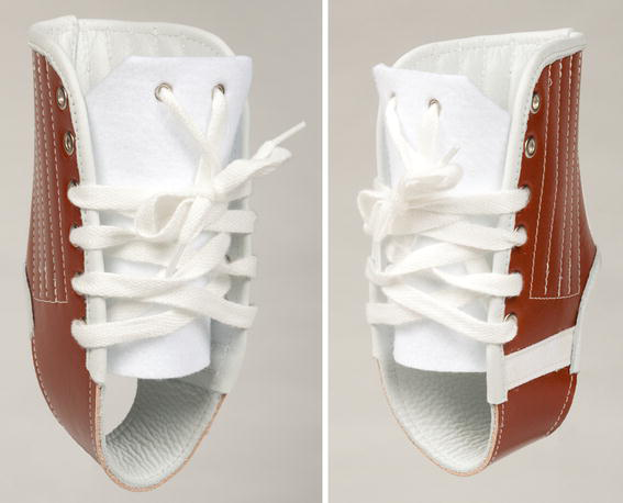
Lace-up gauntlet brace
Of the 100 surgeries performed, 53 cases had at least a 2-year follow-up (average 4 years; range 2–13 years). These cases were evaluated for the following outcomes: return to all activities desired by the patient, ligamentous laxity, pain, and any subsequent sprains. Some limitations to our study include the fact that it was a retrospective report. Forty-seven cases did not have at least a 2-year follow-up. Despite this fact, we still had 53 cases with a 2-year follow-up compared to a previous article with 44 cases [27]. In our opinion, return to activity, postoperative ankle laxity, pain, and subsequent sprains are preferred measures of outcomes. Because articles concerning what the “perfect outcome” is regarding reconstructive ankle surgery are scarce, we consider that the patient returning to activity (including sports) with decreased laxity, pain, and subsequent sprains is a reasonable outcome. Chrisman and Snook describe a satisfactory reconstruction of the lateral ligaments of the ankle joint as the elimination of patient-perceived instability, restoration of function (both occupational and recreational), maintenance of a near-normal range of motion of the ankle in all directions, and lack of sensory changes consequent to surgical manipulation of the sural nerve and its branches, such as sensitivity, numbness, or neuroma formation [28].
Operative technique
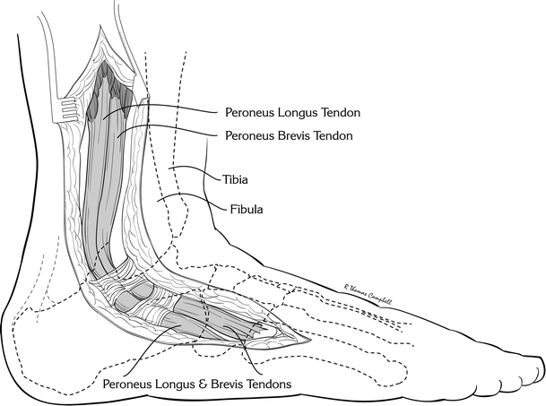
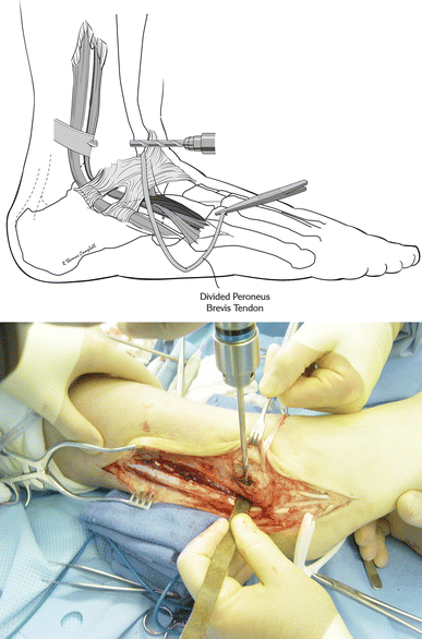
The procedure utilized in this study was adapted from the original Chrisman–Snook procedure, modified by routing the anterior half of the peroneus brevis tendon subperiosteally along the lateral wall of the os calcis during its course back to the site of distal attachment on the fifth metatarsal. The original Chrisman–Snook procedure instead utilized a bone flap in the lateral os calcis, creating a horizontal tunnel through the calcaneus. A long angular incision is made from the mid-calf laterally along the course of the peroneal tendons beneath the lateral malleolus and angling down to the base of the fifth metatarsal (Fig. 2). Any branches of the sural nerve are identified distally and protected throughout the procedure. The sheath of the peroneal tendons is opened and the peroneus brevis and longus identified. The peroneus brevis is tracked distally to its insertion on the base of the fifth metatarsal. Near the level of the ankle, a 1.5 cm section of peroneal sheath is preserved to prevent subluxation. Dissection then proceeds proximally, exposing the tendinous origin of the peroneus brevis by retracting the peroneus longus posteriorly. The anterior half of the peroneus brevis tendon is split longitudinally from its posterior half distally to proximally and resected around mid-calf level (Fig. 3). The tendon itself is cleaned of any muscle fibers. The anterior half of the peroneus brevis tendon is then pulled inferiorly to a site opposite the base of the fifth metatarsal. Next, with a large curved hemostat, the anterior half of the peroneus brevis is passed beneath the sinus tarsi to a site opposite to the anterior portion of the distal fibula about 1.0–1.5 cm above the tibial plafond (Fig. 4). An anterior to posterior cylindrical tunnel, large enough to house the tendon transfer, is then drilled through the distal fibula (Fig. 5). Using a Hewson tendon passer, the tendon is coursed through the distal fibula. After passing the tendon down through the tunnel, the anterior half of the peroneus brevis is brought over the peroneus brevis and longus and then alongside and distally, using a long hemostat, through a subperiosteal tunnel along the lateral wall of the os calcis, eventually reaching the distal attachment on the fifth metatarsal (Fig. 6). With the ankle brought into a position of 90° and slight eversion, the loop of tendon is pulled under tension. The transferred portion of peroneus brevis is then anchored to its posterior half and also to its initially transferred anterior half as it passes underneath the sinus tarsi with multiple interrupted figure-of-eight sutures of 4–0 braided polyester sutures (Fig. 7). A 1–0 chromic suture is used to anchor the tendon as it passes through the fibular tunnel (Fig. 8). The duration of surgery averages roughly 90 min.
Drawing of surgical incision
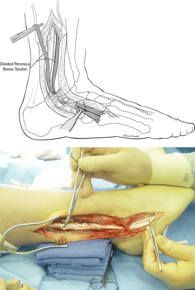
Drawing and operative photograph of peroneus brevis tendon splitting
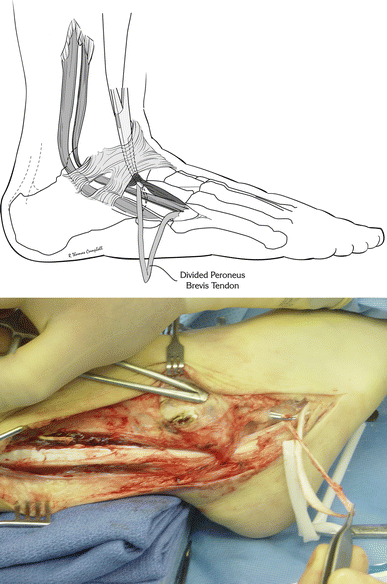
Drawing and operative photograph of tunnel beneath sinus tarsi and extensor digitorum
Drawing and operative photograph of fibular drilling
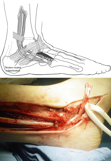
Drawing and operative photograph of split peroneus brevis tendon brought through the lateral subperiosteal tunnel along the lateral wall of the os calcis, eventually reaching the distal attachment on the fifth metatarsal
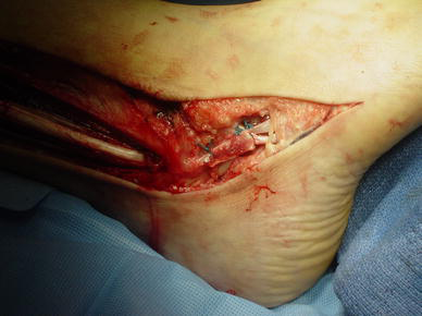
Operative photograph of sutures
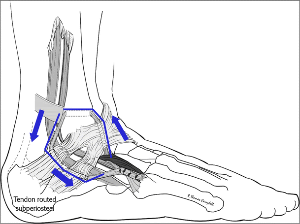
Drawing of complete surgery
Results
Of the 100 surgeries performed, not a single patient developed a deep wound infection or compartment syndrome. There were 10 cases of minor superficial wound healing problems, all of which subsided without incidence. There were two cases of temporary nerve dysfunction, one of which resolved without surgical intervention and the other is still resolving with no plans for surgical intervention. There were two cases of sural nerve entrapment that required surgical treatment due to neuroma formation. One patient with an OCD of the talus prior to the ligamentous laxity surgery did have a subsequent surgery to repair the OCD. There were 87 cases with follow-up greater than 6 months. Not a single patient required subsequent surgery for ankle instability.
Of the 53 cases with at least a 2-year follow-up, all patients returned to full activities of their choice. All cases but one maintained ≤45° of ankle inversion postoperatively. Most ankles (41 cases) demonstrated ≤30° inversion, with the majority in the range of 10°–15° (Table 1). Only one patient developed 70° of ankle inversion but remained asymptomatic and did not require further ankle surgery. All ankles were either totally pain free (39 cases) or experienced only transient discomfort (14 cases) with prolonged running (4 h daily) or standing (8 h at work) (Table 1). Forty-one cases (77%) reported no postoperative sprains; the remaining 12 cases (23%) reported two or fewer minor sprains during the follow-up period (Table 1). In spite of the sprains that occurred to the surgically repaired ankle, no deleterious effects to the repair were made. They were treated routinely as minor ankle sprains.
Characteristics and outcomes of patients with >2 years follow-up
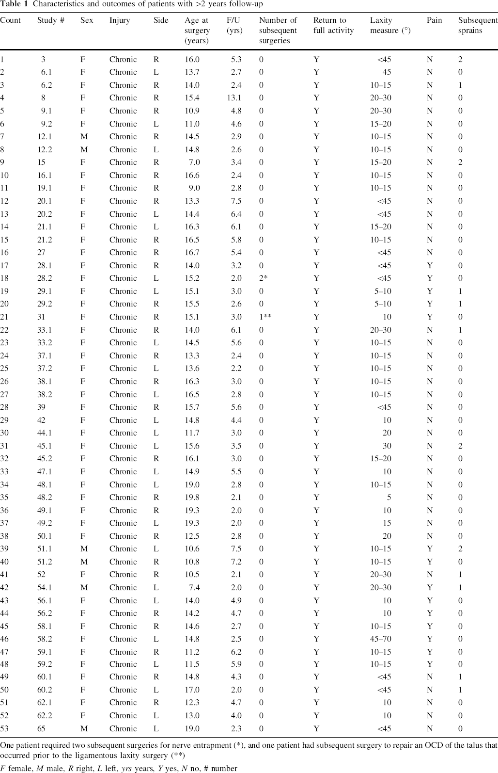
One patient required two subsequent surgeries for nerve entrapment (*), and one patient had subsequent surgery to repair an OCD of the talus that occurred prior to the ligamentous laxity surgery (**)
F female, M male, R right, L left, yrs years, Y yes, N no, # number
Discussion
An increase in chronic ankle instability cases, especially in the pediatric population, has been hypothesized to be secondary to a greater participation of children in organized sports, an increase in motor vehicular accidents, and frequent inadequate treatment of ankle sprains [27]. Treatments of ankle sprains included casting, physical therapy, and bracing when indicated. Cast immobilization in a below-knee walking cast is usually followed by up to 12 weeks of physical therapy [29]. Failure to provide proper initial ankle sprain treatment may result in chronic ligamentous laxity and may lead to a need for surgical intervention.
During adolescence, the ATFL can be disrupted completely with severe supination injury of the ankle, even in the absence of avulsion fractures of the talus or fibula. Many authors have concluded that the ATFL is almost always disrupted in unstable ankles, and in severely unstable ankles the CFL is usually torn. According to Chrisman and Snook, both the talotibial and subtalar joint ligaments need to be reconstructed in patients with disabling instability who wish to engage in heavy work or vigorous athletics [28]. Chronic ligamentous ankle laxity may lead to more long-term consequences of functional instability, recurrent ankle sprains, and degenerative arthritis [30].
The long and slender ATFL is the main stabilizer of the lateral ankle and is the most commonly injured ligament with inversion sprains [21]. Throughout its arc of motion, the ATFL restrains inversion of the ankle. Most tears of the ATFL occur during inversion, plantar flexion, and internal rotation. A positive anterior drawer sign implies damage or rupture of the ATFL [17, 31]. The CFL is situated superficially to the joint capsule beneath the peroneal tendon sheath. Functionally it helps to stabilize the ankle joint along with the subtalar joint, and is injured primarily in ankle dorsiflexion and inversion [10].
Inversion testing can be used to evaluate CFL integrity [9]. The function of the CFL has been a topic of debate ranging from being of limited significance in chronic ankle instability to being the most important lateral stabilizer [32]. The ATFL is ruptured in two-thirds of all acute ankle ligament injuries, and 20% more have simultaneous rupture of the CFL [33]. Isolated CFL rupture is rare, and the PTFL is even more rarely torn with ankle inversion injuries. Recurrent ankle sprains in children, particularly in ligamentously lax individuals, often lead to chronic instability [27].
Ankle sprains are often one of the most poorly managed injuries seen in emergency rooms. Frequently, nonoperative treatment of acute ankle sprains consists of rest, ice, compression wrapping, elevation, and early mobilization. This includes weight bearing at various periods of time, often before edema and ecchymosis subsides. Functional braces are often utilized for patients with chronic sprains [23]. Surgery, indicated by mechanical and functional instability, is generally reserved for those patients who develop recurrent ankle sprains and laxity that are unresponsive to orthotic management.
Some studies have stated that primary anatomic repairs such as the Broström are recommended as the first choice in adults who need full anatomic motion of the ankle in activities such as ballet [21]. Ankle reconstruction with secondary tendon augmentation is preferred in cases of ligamentous laxity, patients with chronic ligamentous insufficiency, and in patients where the Broström ligament repair procedure has failed [10].
Nonaugmented, anatomic repairs such as the Broström and Karlsson have been found to restore normal anatomy and joint mechanics, preserve subtalar motion, yield no donor-site morbidity, and do not change gait mechanics. Disadvantages of this type of repair include unsuccessful stabilization of weak attenuated ligamentous structures seen in chronic ligamentous laxity, and also the failure to address the subtalar instability that occurs with CFL injury [34]. Augmented ankle reconstructions have been described by Evans [14], Watson-Jones [15], Anderson [16], Saltrick [17], Elmslie [18], and Chrisman and Snook [19]. These procedures have been said to lack anatomic ligament reconstruction and, as a result, yield decreased subtalar motion. These claims have been disputed in the literature by White and Kraynick and St. Pierre et al., who state that the peroneus brevis tendon is expendable [35], and ankle reconstruction with the peroneus brevis tendon was found via the Cybex II evaluation to not have significantly weaker eversion when compared to the contralateral normal ankle [36]. As a result, many modifications to these procedures have been created. Hollis et al. reported that inversion–eversion subtalar motion was not significantly changed after augmented reconstruction [22]. Many studies have shown successful results with augmented ankle reconstruction procedures [3, 14, 19, 23, 28, 30, 34, 35, 37–40].
A modified version of the Broström is recommended as the first choice in adults who need full anatomic motion of the ankle for activities such as ballet dancing. This is because of the ability to restore normal anatomy and joint mechanics, along with the above-listed benefits of anatomic reconstruction [21]. Long-term outcome of anatomic ankle reconstruction remains unclear [27]. This is due to the fact that no clear consensus exists as to which procedures produce the best results, and also “excellent end-outcomes” are not clearly defined. For instance, a patient who is not involved in sports may simply define the ability to painlessly return to work as an excellent outcome, while an athlete who can return to work but is no longer performing at his or her former level could rate the surgery as having a poor outcome. Biomechanical studies that support these anatomic repairs evaluated normal cadaver ligaments in vitro and without prior ligamentous laxity [20, 40].
Ankle reconstruction with secondary tendon augmentation is the preferred method in cases of ligamentous laxity, patients with ligamentous insufficiency and chronic ankle sprains, and patients where an anatomic ligament repair procedure has failed [3, 11, 23, 24, 27, 29, 30, 37, 39]. Our patients underwent augmented ankle reconstruction after a protracted history of recurrent sprains and failed bracing attempts. In this study, a failed bracing attempt was defined as recurrent ankle sprains while regularly wearing a lace-up ankle gauntlet for at least 6 weeks; the majority underwent a 6-month trial. In our population of patients, an anatomic reconstruction would more than likely be impossible due to the extreme excessive laxity of the ligamentous tissue. In as much as there are no long-term studies available in the pediatric population, it is our current opinion that the modified Chrisman–Snook repair offers a distinct advantage in this group of ligamentously lax individuals.
Our modification of the Chrisman–Snook operative procedure routinely results in a stable ankle with a significant reduction in the range of inversion. Patients were able to return to full activities on average 4 months after surgery. Of the 53 cases with at least a 2-year follow-up, all patients returned to full activity of their choice.
Sural nerve injury is important to note, and careful dissection of its branches is important in preventing this complication. Upon identification, the sural nerve is surrounded and protected throughout the procedure. Numbness and paresthesia over the lateral aspect of the foot and ankle may result from sural nerve injury [30].
Although preoperative plain radiographs were obtained in all patients, there are very poor correlations between radiographic definitions of instability, and variations in technique have led many to rely on the clinical exam alone [3]. If radiography is to be useful in the future assessment of instability, a more precise method is needed to evaluate both talocrural joint and subtalar joint movement [41].
Conclusion
The author's modification of the Chrisman–Snook procedure as described in this study has been successfully employed in 100 consecutive cases of chronic ligamentous instability in children and adolescents with excellent overall results and exceedingly few complications. All patients were able to return to all desired activities of daily life.
Footnotes
Acknowledgments
The authors wish to express their appreciation to Tom Campbell, medical illustrator, for his drawings, and Martin S. Dicintio, assistant research coordinator at Akron Children's Hospital, for his copyediting and manuscript preparation.
None of the authors received financial support for this study; nor are there any potential, perceived, or real conflicts of interest. The study was approved by the institutional review board at Akron Children's Hospital.