
Abstract
A nanosilver composite plant medium with a good antimicrobial capability was successfully prepared via a two-step method in this work. First, nanosilver particles were prepared in a silver nitrate (AgNO3) solution by using D-glucose as a reduction reagent at room temperature. The effect of AgNO3 concentration levels on the size of the nanosilver particles was investigated. The sedimentation activation energy of the nanosilver particles was also measured. Second, the solvent containing the nanosilver particles was added to a Murashige-Skoog medium (a plant growth medium used in the laboratory for the cultivation of plant cell cultures, which was invented by Toshio Murashige and Folke K. Skoog) to form a composite plant medium. The antimicrobial capability of the composite medium was investigated by cultivating in it some fresh strawberry slices. The results showed that the concentration level of AgNO3 significantly influenced the average size of the prepared nanosilver particles. When the AgNO3 concentration was increased from 1.0×10⁻³ mol/L to 4.0×10⁻³ mol/L, the average diameter of the nanosilver particles increased from 52 nm to 72 nm, and the sedimentation activation energy of the particles increased from −20.0 kJ/mol to −63.3 kJ/mol. Images from the transmission electron microscope showed that the size distribution of the particles was within narrow parameters. The results of sedimentation activation energy strongly supported the size results obtained from the TEM measurement. It meant that the preparation method could offer good control over the size and size distribution of the nanosilver particles. The cultivation experiments proved that the composite medium has a good antimicrobial capability. Since D-glucose is a nontoxic material, this method of preparing the composite MS medium with an antimicrobial capability was a ‘green’ method.
1. Introduction
Contamination is a common problem in tissue cultures [1]. In the process of developing a traditional plant tissue culture, the bacteria carried or bred using plants are hard to be cleared, and are brought to the medium. In addition, the sterilization of the culture medium and workbench is not stringent or the sterilization operation is nonstandard in the process of plant breeding. This leads to further pollution, consequently slowing the traditional tissue culture plant growth and decreasing the survival rate. These problems have inhibited the use of plant tissue culture in actual agricultural production and promotion [1-3].
If plants can grow under relatively open conditions, then investments in manpower and material resources would be reduced. In addition, this condition would also be beneficial for the promotion of plant tissue culture technology being used widely in the commercial production of young plants. However, in practice, open tissue culture technology has no resistance to mixed bacteria in the air. Thus, the addition of antibacterial agents to the medium would be beneficial for open tissue culture.
Nanosilver has natural antibacterial properties. Silver nanoparticles can kill hundreds of various types of bacteria in minutes. Jayaprakash and Li [4, 5] revealed that silver nanoparticles could inhibit the growth of Escherichia coli, S. aureus, C. albicans, B. cereus, M. luteus, Enterococcus, P. aeruginosa and other pathogenic microorganisms. Therefore, the addition of nanosilver particles into the plant medium would allow the composite medium to possess antimicrobial capabilities.
Silver ions are usually reduced by using reduction reagents in the solution to obtain nanosilver particles. The common reduction reagents used are formamide, pentanediol, hydrazine, triethylamine, and polyvinylpyrrolidone, among others [6-11]. In addition, a number of ‘green’ methods using some nontoxic natural materials (e.g., D-glucose, starch and fructose) as reduction reagents were also invented [12,13]. However, for many methods of preparing nanosilver particles, a common disadvantage was the inability to control the size (including size distribution) of the particles [13].
In this paper, in order to prepare a plant medium with an antimicrobial capability, nanosilver particles were first obtained by reducing silver nitrate with D-glucose, which could also be used as energy for the plant medium. Compared with using the common organic solvents as reduction reagents, using D-glucose is a ‘green’ method. The effect of silver ion concentration on the size of the nanosilver particles was researched and the sedimentation activation energy of the nanosilver particles was also measured. It was found that the size of the prepared nanosilver particles could be controlled easily by adjusting the solution's silver ion concentration. In addition, the solvent containing the nanosilver particles was then added to a Murashige-Skoog (MS) medium (a plant growth medium used in the laboratory for the cultivation of a plant cell culture, which was invented by Toshio Murashige and Folke K. Skoog in 1962) to form a composite plant medium. The antimicrobial capability of the composite plant medium was also investigated.
2. Materials and Methods
2.1 Materials
Silver nitrate (AgNO3, analytic reagent) was purchased from Beijing Jingxi Chemical Co. Ltd., China. Ammonium hydroxide (NH4OH, analytic reagent) and sodium hydroxide (NaOH, analytic reagent) were purchased from Tianjin Kermel Chemical Reagent Development Center, China. An MS medium (biological reagent) containing sucrose and agar was purchased from Linan Mumu Biological Technology Co. Ltd., China. D-glucose (guaranteed reagent) was purchased from Sigma-Aldrich Biotechnology Company. All reagents were used directly, without further purification.
2.2 Preparation of nanosilver particles
In a dimly lit laboratory, four kinds of nanosilver particles with different sizes were prepared in 50 mL Erlenmeyer flasks via the ‘green’ and nontoxic method at four AgNO3 concentrations of 1.0×10⁻³ mol/L, 2.0×10⁻³ mol/L, 3.0×10⁻³ mol/L and 4.0×10⁻³ mol/L, using D-glucose as a reduction reagent at room temperature. First, 5 mL of 0.005 mol/L NH4OH was added to 20 mL of AgNO3 solution. The mixture was then stirred using a magnetic agitator for two minutes at room temperature. Afterwards, 10 mL of 0.015 mol/L D-glucose solution was added to the mixture and the pH value of the solution was adjusted to 9.0 by adding a few drops of 0.01 mol/L NaOH solution. Finally, the obtained solution was kept in place at room temperature for 1 h. The colour of the reacting solution turned from colourless to yellow.
2.3 Characterization of nanosilver particles
The four solutions containing nanosilver particles were characterized using a UV-vis spectrophotometer (T6, Persee, Beijing, China) to measure the wavelength (λmax) and absorbance (Imax) of the maximum absorption peaks. The morphology of the obtained nanosilver particles was observed under a transmission electron microscope (FEI Tecnai F20, Hillsboro, Oregon, USA) with 200 kV acceleration voltage. The average diameter of the particles was determined from the TEM photos. In addition, the elemental composition of the particles was also analysed by using the EDS (Energy-dispersive X-ray spectroscopy) function of the TEM equipment in order to confirm the formation of silver particles rather than other particles.
2.4 Measurement of the sedimentation activation energy of nanosilver particles
The prepared nanosilver particles would begin to slowly sediment in the solutions when no stabilizing agents were added. The sedimentation performance of the particles in the solutions after resting for 72 h was measured at 15°C, 25°C, 30°C and 40°C. First, the initial absorbance Imax-0 (at λmax) of the solutions was measured using the UV-vis spectrophotometer when nanosilver particles formed in the solutions. After resting for 72 h, the absorbance Imax-72 (at λmax) of the upper solutions in the Erlenmeyer flasks was measured using the UV-vis spectrophotometer. The relative change (k) of absorbance was calculated via the following equation:

The sedimentation activation energy Ea was calculated using the principle of Arrhenius' equation as follows:

where A is a pre-exponential factor, R is the universal gas constant and T is the absolute temperature. When plotting ln k versus 1/T, a line would be obtained. Then Ea could be calculated from the slope of the line.
2.5 Test of the antimicrobial capability of a nanosilver composite plant medium
To test the antimicrobial capability of the composite medium, the nanosilver particles reduced in the concentration of 1.0×10⁻³ mol/L AgNO3 were chosen to prepare a composite medium. First, 4.2 g MS medium powder was dissolved in 100 mL of sterile water. The mixture was then heated to completely dissolve the MS medium. Up to 10 mL of the solution containing the above nanosilver particles was then added to the medium solution. After stirring, the nanosilver composite plant medium was obtained and transferred to a culture dish.
Fresh strawberry slices were cultivated in the medium containing the nanosilver particles. For comparison, the medium without the addition of nanosilver particles was also used to cultivate fresh strawberry slices. The two media were cultivated in air without sterilization. The growth state of the mixed bacteria in the two media was observed.
3. Results and discussions
3.1 Morphology and UV-vis absorption of nanosilver particles
For a solution containing nanosilver particles, the UV-vis absorption spectra of the solution will present a dominant absorption peak at about 400∼500 nm [10,14-19]. Figure 1 presents the UV-vis absorption spectra (from 300 nm to 800 nm) of the four solutions containing the nanosilver particles. A dominant absorption peak was observed between 440 and 540 nm for each curve, indicating the presence of the nanosilver particles. The figure also shows that the position of the absorption peak moves toward the longer wavelength with the increase in AgNO3 concentration from 1.0×10⁻³ mol/L to 4.0×10⁻³ mol/L. Moreover, the size of the nanosilver particles increased with a redshift of the UV-vis absorption spectrum [10, 15-18]. The width of the absorption spectrum can reflect the size distribution of the nanosilver particles. A 4.0×10⁻³ mol/L AgNO3 solution had the widest absorption peak, indicating that the size distribution of the nanosilver particles in this solution was the largest.

UV-vis absorption spectra of solutions containing nanosilver particles
Figure 2 presents the TEM images of the nanosilver particles prepared in the four AgNO3 solutions. It shows that most of the nanosilver particles are near spherical particles with good dispersibility and that their size distribution is not large. The average diameter of the nanosilver particles is 52 (a), 58 (b), 65 (c) and 72 (d) nm, respectively. Therefore, the average diameter of the nanosilver particles increases with an increase in AgNO3 concentration.

TEM images of nanosilver particles prepared in AgNO3 solution: (a) 1.0×10⁻³ mol/L AgNO3, (b) 2.0×10⁻³ mol/L AgNO3, (c) 3.0×10⁻³ mol/L AgNO3 and (d) 4.0×10⁻³ mol/L AgNO3
Figure 3 presents the results of EDS elemental analysis of the nanoparticles prepared in 3.0×10⁻³ mol/L AgNO3 solution. The peaks of the copper element came from the copper net used to support the sample in the TEM column. The peak of the silicon element came from the electron gun of the TEM equipment. All of the other peaks belong to the silver element. Consequently, the prepared nanoparticles were silver nanoparticles.
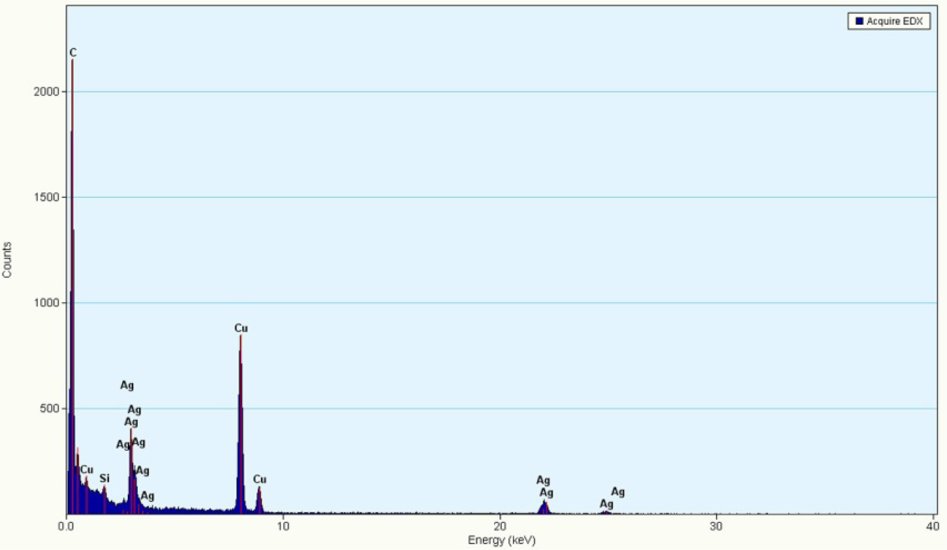
EDS elemental analysis of nanoparticles prepared in 3.0×10⁻³ mol/L AgNO3 solution
Figure 4 presents the plots of the average diameter of nanosilver particles versus the wavelength (λmax) of the UV-vis maximum absorption peak and the corresponding absorbance (Imax). It shows clearly that λmax increases linearly with the increase in average diameter, but that Imax decreases with the average diameter.

Plots of the average diameter of nanosilver particles versus wavelength of UV-vis maximum absorption peak and absorbance
3.2 Sedimentation activation energy of nanosilver particles
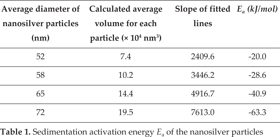
Figure 5 presents the plots of ln k versus 1/T of the four solutions containing nanosilver particles. For each solution, the relative change k of the absorbance was calculated using Eq. (1). As shown in this figure, there is always an approximately linear relationship existing for the four dots of each solution. Table 1 lists the slope values of the four fitted lines shown in Figure 5. The larger slope value indicates a larger sedimentation speed. For the nanosilver particles 72 nm in size, the sedimentation speed was about three times that of the sedimentation speed of 52 nm particles. The sedimentation activation energy Ea of each kind of nanosilver particle was then calculated using Eq. (2), and the final results are also listed in Table 1.
Sedimentation activation energy Ea of the nanosilver particles

Plots of logarithmic k (relative change of absorbance) versus the reciprocal of temperature: (▪) 1.0×10⁻³ mol/L AgNO3, (•) 2.0×10⁻³ mol/L AgNO3, (▴) 3.0×10⁻³ mol/L AgNO3 and (▾) 4.0×10⁻³ mol/L AgNO3
Figure 6 shows the plot of the sedimentation activation energy Ea, versus the average diameter of the nanosilver particles. It presents clearly the finding that with the increase in average diameter, more sedimentation activation energy was needed (the negative values mean that the sedimentation activation energy was exothermic). As shown in Table 1, for the 72 nm particle and the 52 nm particle, the calculated average volume increased by about three times. The sedimentation activation energy also increased by about three times. Therefore, the consistent results above prove that the measured average diameter values using TEM images were precise.

Plot of sedimentation activation energy versus average diameter of nanosilver particles
3.3 Test of antimicrobial activity
Figure 7 shows the images of two kinds of MS media. One was without nanosilver particles and the other contained nanosilver particles. The MS medium containing nanosilver particles presents as yellow. Figure 8 shows the images of strawberry slices cultivated in the two MS media in air. After three days, mould appeared on the surface of the MS medium without nanosilver particles, surrounding the strawberry. However, no mould was observed on the surface of the composite MS medium containing nanosilver particles. On the fifth day, the quantity of mould on the pure MS medium increased, but still no mould was observed in the composite MS medium. The mould breeding rate was significantly influenced by the antibacterial properties of its environment. In a medium containing an antibacterial substance, such as antibiotics, ethanol or silver, etc., mould breeding would be strongly inhibited. Therefore, for the cultivation experiments described above, the results demonstrated that the culture medium containing the nanosilver particles had a significant antibacterial capability.

Images of MS media: (a) MS medium and (b) composite MS medium containing nanosilver particles
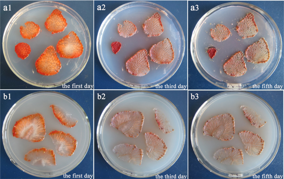
Images of strawberry slices cultivated in two kinds of MS media: (a) MS medium and (b) composite MS medium containing nanosilver particles
4. Conclusions
First, nanosilver particles were successfully prepared by reducing silver nitrate with D-glucose. When the AgNO3 concentration was increased from 1.0×10⁻³ mol/L to 4.0×10⁻³ mol/L, the average diameter of the nanosilver particles increased from 52 nm to 72 nm and the sedimentation activation energy of the particles increased from −20.0 kJ/mol to −63.3 kJ/mol. Images from the transmission electron microscope showed that the size distribution of the particles was within narrow parameters. The results of sedimentation activation energy strongly supported the size results obtained via TEM measurement. This meant that the preparation method might offer good control over the size and size distribution of the nanosilver particles.
Second, the composite MS medium containing the nanosilver particles was also successfully prepared. The cultivation experiments using strawberry slices as samples proved that the composite medium had a good antimicrobial capability. Due to D-glucose being a nontoxic material, this method of preparing the composite MS medium with an antimicrobial capability was a ‘green’ one.
Footnotes
5. Acknowledgements
The authors acknowledge the financial support of the Science and Technology Planning Project of AnKang University (2015AYPYZR01) and the National Natural Science Foundation of China (21376048).