
Abstract
Silver molybdate (Ag2MoO4) and silver tungstate (Ag2WO4) nanomaterials were prepared using two complementary methods, microwave assisted hydrothermal synthesis (MAH) (pH 7, 140 °C) and co-precipitation (pH 4, 70 °C), and were then used to prepare two core/shell composites, namely α-Ag2WO4/β-Ag2MoO4 (MAH, pH 4, 140 °C) and β-Ag2MoO4/β-Ag2WO4 (co-precipitation, pH 4, 70 °C). The shape and size of the microcrystals were observed by field emission scanning electron microscopy (FE-SEM), different morphologies such as balls and nanorods. These powders were characterized by X-ray powder diffraction and UV-vis (diffuse reflectance and photoluminescence). X-ray diffraction patterns showed that the Ag2MoO4 samples obtained by the two methods were single-phased and belonged to the β-Ag2MoO4 structure (spinel type). In contrast, the Ag2WO4 obtained in the two syntheses were structurally different: MAH exhibited the well-known tetrameric stable structure β-Ag2WO4, while co-precipitation afforded the metastable β-Ag2WO4 allotrope, coexisting with a weak amount of the α;-phase. The optical gap of β-Ag2WO4 (3.3 eV) was evaluated for the first time. In contrast to β-Ag2MoO4/β-Ag2WO4, the α-Ag2WO4/β-Ag2MoO4 exhibited strongly-enhanced photoluminescence in the low-energy band (650 nm), tentatively explained by the creation of a large density of local defects (distortions) at the core-shell interface, due to the presence of two different types of MOx polyhedra in the two structures.
1. Introduction
Tungstate ceramic materials are mixed oxides that can be used in numerous technological applications such as humidity sensors [1], photocatalysts [2] electrochromic devices [3], Raman converters [4], scintillators [5], photoluminescent materials [6] and lasers [7–8]. α-Ag2WO4 can be considered an example of a high-dimensional and high-connected polyoxometalate-based hybrid compound [9–10] Pang et al. prepared superfine Ag2WO4 antibacterial powders with good antimicrobial durability [11] and using α-Ag2WO4 as a starting material, prepared an Ag/AgBr/WO3.H2O composite that exhibited good photocatalytic activity for the destruction of Escherichia coli bacteria under visible light and that was relatively stable under repeated use. [12]. Pan et al. [13] synthesized microstructured Ag2WO4 with a shuttle-like shape via a precipitation process with the assistance of Arabic gum. Aouadi et al. [14] studied the changes in the chemistry and crystal structure of Ag2Mo2O7 and Ag2WO4 thin films and powders ranging between 25° and 600° C. In addition, they used ab initio molecular dynamics and different experimental techniques to explain the low coefficient of friction.
L.S. Cavalcante et al. observed and explained the photoluminescent behaviour of α-Ag2WO4 (i.e., Ag8W4O16) microcrystals [15], in which Ag ions can display different AgOx corresponding to type-coordination and synergistic coordination modes in order to facilitate the formation of several clusters with low and high coordination numbers (x=2,4,6,7). The latter phase, which is the thermodynamically stable phase among several polymorphs, namely α-, β-, and gamma;- Ag2WO4 [16], has been found to be an interesting material, due to the growth of Ag filaments on its surface when it is exposed to electron beam irradiation [17]. Recently, controlled synthesis and the high photocatalytic activity of hierarchically porous metastable β-Ag2WO4 hollow nanospheres were reported by Wang et al. [18].
Consequently, silver tungstate Ag2WO4 has been receiving attention from the scientific community due to its vast scope for applicability. Its photoluminescent properties [19] and corresponding applications as a microbial agent [20], as well as its use as a novel ozone gas sensor [21], were investigated by our group.
Metal molybdates, another important family of inorganic materials, have also been intensively investigated because of their various properties, one of these being photoluminescence [22–24]. Several studies have been carried out to explore the properties and practical applications of silver molybdate Ag2MoO4, which exist in two forms, α-tetragonal [25] and β- cubic [26–27]. Silver molybdates have applications in areas such as high-temperature lubrication [28–29], ion-conducting glasses [30] and gas-sensing and surface enhanced Raman scattering techniques [31]. To date, flower-like [32] nanoparticles [33] and wire-like [34] have been obtained Tai et al. [35] reported the MAH synthesis of cube-like Ag-Ag2MoO4, which has visible-light photocatalytic activity. Low- dimensional silver molybdate-nanostructures have been obtained by Nagaraju et al., who reported the synthesis of nanorods/nanowires/multipods and the photoluminescence of microrods [36]. Lei et al. [37] reported on the room-temperature synthesis of Ag nanoparticles with decorated silver molybdate nanowires, using a solution-based chemical reaction method. Long et al. [31] published a facile hydrothermal technique for synthesizing Ag2MoO4 at relatively low temperatures (
Three more sections complete this article. Next, we present the experimental details. In Section 3, the results are presented and discussed. A brief summary and the main conclusions will be offered in the final section.
2. Experimental Details
2.1 Synthesis of Ag2WO4, Ag2MoO4 and decorated nanoparticles
Two sets of samples were synthesized, with and without the use of MAH synthesis.
In the first set, silver tungstate was precipitated by mixing solutions of Na2WO4.2H2O and AgNO3 (1 mmol and 2 mmol, respectively, in 50 ml of deionized water). The suspension was transferred in a PTFE autoclave and heated at 140 °C for 60 min with microwave assistance (2.45 GHz, maximum power of 800 W). Then, the precipitate was separated by centrifugation, washed several times with deionized water and ethanol and dried at 70 °C for 24 hours. For reference, silver molybdate was prepared following the same procedure. In a second step, 0.2 g of silver tungstate was added to 50 mL of distilled H2O and dispersed by ultrasonic agitation for 10 min. To this solution, 25 mL (1 mmol) of sodium molybdate and 25 mL (2 mmol) of silver nitrate were added under vigorous magnetic stirring. The pH of the solution was adjusted to 4 by the addition of nitric acid and the mixture was, as previously, heated at 140 °C for 60 min with microwave assistance. Separation and drying were applied as above. In the following, the two obtained compounds will be labelled as W-htmw and Mo-htmw, and the silver tungstate nanoparticles decorated by the silver molybdate as W/Mo-htmw.
For the second set, silver tungstate and silver molybdate were prepared by precipitation from sodium salts in aqueous solutions of pH 4 (nitric acid and distilled water). For silver tungstate, 1 mmol of Na2WO4.2H2O (99.5% purity, Sigma Aldrich) was dissolved in 50 ml of pH 4 solution (solution A). Separately, 2 mmol of AgNO3 (99.8% purity, Sigma Aldrich) was dissolved in another 50 ml of the pH 4 aqueous solution (solution B). These two solutions were heated at 70 °C; then, solution B was added to solution A under vigorous magnetic stirring, after which the solution became dark grey. The mixture was kept at 70 °C for 10 min with stirring. Then, the precipitate was separated by centrifugation, washed several times with deionized water and ethanol, and dried at 70 °C for 24 hours. Silver molybdate was prepared following the same procedure using Na2MoO4.2H2O (99.5% purity, Sigma Aldrich). In the following, the obtained compounds will be labelled as W-70 and Mo-70, respectively. To prepare silver molybdate nanoparticles decorated by the silver tungstate, 0.2 gr of sample Mo-70 was added to 50 ml of the pH 4 solution and dispersed for 15 min by ultrasonic agitation. Then, 1 mmol of Na2WO4.2H2O and 2 mmol of AgNO3 were dissolved separately in 25 ml of ph 4 solution (solutions 1, 2 and 3, respectively). The three solutions were heated at 70 °C and then solutions 2 and 3 were added to solution 1 sequentially and kept under agitation for 10 min. The silver tungstate nanoparticles decorated by the silver molybdate were prepared in a similar manner. The decorated samples obtained were labelled Mo/W-70 and W/Mo-70.
2.2 Characterization of Ag2WO4, Ag2MoO4 and decorated nanoparticles
The Ag2WO4 and Ag2MoO4 microcrystals and the decorated nanoparticles were characterized by X-ray diffraction (XRD) patterns, using a D/Max-2000PC diffractometer Rigaku (Japan) with Cu Kα radiation (λ = 1.5406 Å) in the 2θ range from 10° to 70° in the normal routine and with a scanning velocity of 2°/min. The shapes and sizes of the Ag2WO4 and Ag2MoO4 microcrystals and decorated nanoparticles were observed with a FE-SEM model Inspect F50 (FEI Company, Hillsboro, OR), operated at 5 kV. UV-vis spectra were taken using a Varian spectrophotometer (model Cary 5G) in diffuse-reflectance mode. PL measurements were performed through a Monospec 27 monochromator (Thermal Jarrel Ash) coupled to a R446 photomultiplier (Hamamatsu Photonics, Japan). A krypton-ion laser (Coherent Innova 90K; λ = 350.7 nm) was used as the excitation source; its maximum output power was maintained at 500 mW. The laser beam was passed through an optical chopper and its maximum power on the sample was maintained at 40 mW. All PL measurements were performed at room temperature.
3. Results
3.1 X-ray diffraction
Figure 1 shows the powder XRD patterns of our silver molybdate (Mo-htmw) and silver tungstate (W-htmw) samples, as well as the synthesized composite (W/Mo-htmw) of the first set. It is obvious that the molybdate sample Mohtmw was pure β-Ag2MoO4, as the pattern perfectly matched the JCPDS card no. 08-0473. This result supports previous reports and showed that this phase usually formed near pH 7 [32,40,43]. Similarly, the pattern of our silver tungstate W-htmw exactly matched the JCPDS card no. 34-0061, implying that the sample was perfectly monophased α-Ag2WO4, in good agreement with previous observations in neutral [15,44–46] or slightly alkaline [47] media. The profile of the W/Mo-htmw composite was essentially the superimposition of the patterns of the two components.

X-ray powder patterns of: a) β-Ag2MoO4 (Mo-htmw); b) α-Ag2WO4 (W-htmw); c) α-Ag2WO4/β-Ag2MoO4 (W/Mo-htmw)
Only some exaltation of the intensity of the 131st peak of the tungstate should be mentioned (for which any orientation effect was ruled out because its harmonic was not affected). It is worth noting that the W/Mo-htmw was prepared at pH 4, as experiments at pH 7 failed. This observation prompted the use of pH 4 in the second set of syntheses.
The powder XRD patterns of the samples prepared at atmospheric pressure (pH 4) without the assistance of additional energy are given in Figure 2. Mo-70 appears again as pure spinel β-Ag2MoO4, although this phase is commonly obtained in neutral or alkaline media, as mentioned above. In contrast, the obtained silver tungstate is a mixture of the two orthorhombic α- and hexagonal β- allotropes, where the latter is strongly dominant. This result was actually unexpected, as the stable phase is the α- phase and was systematically obtained by various methods [17,22,40,44–46], including the “coprecipitation” approach proposed by Calvacante [15]. Indeed, the β-phase was previously obtained only by precipitation in ice water at pH 9 [16], or as metastable hollow spheres, using PMMA cross-linked micelles as templates [18]. The choice of temperature and pH appears crucial, as any change leads to the alpha;-allotrope, as seen in the literature data. As expected, the decorated Mo/W-70 exhibited superimposition of the patterns of the two precursors. In contrast, in the case of W/Mo-70 (not shown) it appeared that the β-Ag2WO4 was almost fully transformed in the stable α-phase, due to a longer time at 70 °C, but in addition, significant decomposition occurred, as indicated by the emergence of Ag2O peaks; this sample was not studied further.

X-ray powder patterns of: a) β-Ag2WO4 (W-70); b) β-Ag2MoO4 (Mo-70); c) β-Ag2MoO4/β-Ag2WO4 (Mo/W-70)
3.2 Microstructure
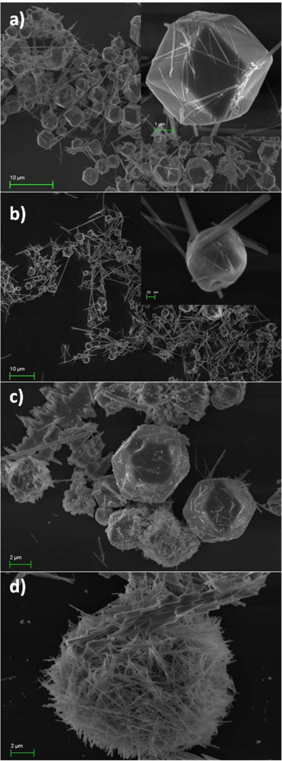
The microstructure of the as-prepared and the composite compounds was revealed by FE-SEM images. Figure 3 displays the photographs of the -htmw samples. As can be seen, Mo-htmw was crystallized in the form of short nanorods with a homogeneous size (200 nm by 1 μm) (Figure 3a) and W-htmw appeared in the form of microsized irregular polyhedral crystals (Figure 3b). The latter were accompanied by a few microribbons that were thought to be the same α-Ag2WO4 phase on the basis of XRD data. W/Mo-htmw images showed mainly micrometric balls exhibiting very compact coverage by platelets, about 200 nm wide (Figure 3, c and d). In addition, some ribbons looked similar to those present in the starting W-htmw, but they were not isolated separately and appeared to have been partly dissolved, probably by an Ostwald ripening mechanism.

SEM image of: a) β-Ag2MoO4 (Mo-htmw); b) α-Ag2WO4 (W-htmw); c) and d) decorated nanoparticles α-Ag2WO4/β-Ag2MoO4 (W/Mo-htmw)
The microstructures of the -70 samples are shown in Figure 4. In contrast to Mo-htmw, the Mo-70 sample showed crystals of two different types Figure 4a): nanowires several dozen Mm long and 100–200 nm in diameter, which closely resembled the monoclinic Ag2Mo2O7 fibres often obtained in hydrothermal experiments carried out at pH 4 [32,40,43].
SEM image of: a) β-Ag2MoO4 (Mo-70); b) α- and β-Ag2WO4 (W-70); c) and d) decorated nanoparticles β-Ag2MoO4/β-Ag2WO4 (Mo/W-70)
However, from the XRD data, the formation of this silver dimolybdate was ruled out. These nanowires coexisted with well-shaped cubic or cuboctahedral microcrystals, almost monodisperse in size (about 3 pm) and contrasting with the irregular nanoparticles usually observed for hydrothermally grown (at a pH of about 7–8) Ag2MoO4 samples [32,39–40,43]. Although exhibiting very different morphologies, these two types of crystals clearly belonged to the same β-Ag2MoO4 phase. as proven by the XRD data.
Concerning W-70, FE-SEM micrographs also showed two types of crystals, i.e., nanowires and polyhedral microcrystals (Figure 4b). In this case, because XRD data clearly showed that the sample contained the two phases α- and β-Ag2WO4, it would be reasonable to attribute the quasispherical crystals to the minor phase α- of the orthorhombic structure and the nanowires to the hexagonal β-phase.
On the FE-SEM micrograph of Mo/W-70, one can observe a number of spherical features, including the sea urchin-like surface, which is made of interpenetrated nanowires forming a continuous, felted coating (Figure 4, c and d). These microspheres appeared to be on average slightly larger than the crystals previously attributed to Mo-70, suggesting that W-70 nanowires were grown on the latter crystals. This coverage was not homogeneous for all Mo-70 cuboctahedral microcrystals, because some of them had compact coverage, while others were only partly covered. In addition, some free-standing nanowires were observed; however, it could not be determined whether they consisted of unmodified remaining Mo-70, or free W-70 nanowires newly formed in solution during the final step of the synthesis.
3.3 Optical absorption
The UV-visible diffuse reflectance spectra of Mo-70, W-70 and composites Mo/W-70 and W/Mo-htmw were recorded and the optical band-gap energy (Eg) was calculated using the method of Kubelka and Munk-Aussig [48]. As seen in Figure 5, for these four examples, a straight line was found when plotting [F(R)hv]2, meaning an exponent n = ½ in the classical formula αhv = C(hv - Eg) n , which is the signature of a direct band-gap. There are only limited data in the literature concerning the band-gaps of molybdates and - tungstates with monovalent counter cations. Z.Q. Li reported an indirect gap of 3.37 eV for a β-Ag2MoO4 sample prepared at 180 °C by microwave assisted hydrothermal synthesis [49], while for Ag2Mo2O7, quite different values of 2.94 [50] and 2.65 eV (indirect) [51] were reported. Concerning the tungstates, Cavalcante et al. measured α-Ag2WO4 values between 3.19 and 3.23 (direct gap) [15], depending on the method of preparation, which were close to an independent estimation of 3.10 [52]. A theoretical calculation afforded a slightly larger value of 3.55 eV [17]. However, the compounds reported here belong (at least, primarily) to the β-Ag2WO4 allotrope, a definitively different structure.

UV-vis spectra of β-Ag2MoO4 (Mo-70), β-Ag2WO4 (W-70), β-Ag2MoO4/β-Ag2WO4 (Mo/W-70) and α-Ag2WO4/β-Ag2MoO4 (W/Mo-htmw)
3.4 Photoluminescence
The photoluminescence of AMoO4 and AWO4 compounds has been largely documented, especially in examples where A is a divalent cation, such as Pb or an alkaline earth [22–24,40,53–58]. As a general rule, multiphonon processes occur i.e., a system in which relaxation occurs by several paths, involving the participation of numerous states within the band gap of the material, leading to several emission bands. It is well known that the high energy component of the PL emission is an emission mainly attributed to charge-transfer transitions within the (MO4)2- tetrahedral groups [PLg,h] or the WO6 octahedra [15]. There is consensus about attributing the lowest energy emission bands to the creation of additional deep electronic levels in the forbidden band gap, relative to defects that can be either geometrical distortions of metal oxygen polyhedra and/or oxygen vacancies [15,23,40,55–58]. This model was clearly confirmed by first-principle calculations [55].
The PL spectra of Mo-htmw, W-htmw and W/Mo-htmw are shown in Figure 6. The three compounds exhibited a weak emission around 450 nm, obviously related to charge transfer transition within regular MoO4 tetrahedra and WO6 octahedra, respectively, as mentioned above. The PL spectrum of W/Mo-htmw presented a strong emission around 650 nm, while the two other compounds showed no significant emission in this wavelength range. The enhancement of the PL spectrum for the decorated sample can be explained by the presence of the Ag2MoO4 shell covering the surface of α-Ag2WO4 core, forming some structural defects at the interface and favouring the enhancement of the PL emission. Indeed, β-Ag2MoO4 and α-Ag2WO4 were built from MoO4 tetrahedra and WO6 octahedra, respectively; consequently, a structural distortion at the surface/interface region between these two compounds cannot be ruled out.

PL emission spectra of β-Ag2MoO4 (Mo-htmw), β-Ag2MoO4 (Mo-htmw) and α-Ag2WO4/β-Ag2MoO4 (W/Mo-htmw)
The samples synthesized at 70 °C without microwave assistance (Figure 7) behaved very differently from those synthesized on microwave: Mo-70 exhibited a strong PL peak around 450 nm, originating from the charge transfer in levels very close to valence and conduction bands, and only a weak contribution near 650 nm, reflecting an ordered structure. This is in contrast to W-70, which is essentially a metastable phase (the β- allotrope, as shown by XRD). In the decorated Mo/W-70, there was also an enhancement of the long wavelength contribution, although less pronounced than in W/Mo-htmw. Although the actual structure of β-Ag2WO4 is still unknown, there is some reason to speculate that it is built from (WO4)2- tetrahedra [PLk]. As a result, at the interface, the polyhedra should be less distorted. Another finding was the decrease of the intensity of the short wavelength PL peak of the molybdate core: this is indicative of good coverage by the tungstate shell.

PL emission spectra of β-Ag2MoO4 (Mo-70); b) β-Ag2WO4 (W-70), β-Ag2MoO4/β-Ag2WO4 (Mo/W-70) and α-Ag2WO4/β-Ag2MoO4 (W/Mo-htmw) for scaling
4. Conclusions
Metal molybdates and tungstates are a family of multicomponent metal oxide compounds that have been extensively studied by the scientific community as multifunctional materials, due to their interesting structures, intriguing physical-chemical properties and their diverse and prominent applications. In this study, we grew nanoparticles of Ag2MoO4 and Ag2WO4 using two complementary techniques: the MAH synthesis at 140 °C and pH 7, and a simple, co-precipitation at pH 4. The two approaches afforded the cubic β-Ag2MoO4; for the silver tungstate, however, the outcome was a very different result. The stable orthorhombic α-Ag2WO4 (i.e., Ag8W4O16 built from tetrameric species of four edge-sharing distorted WO6 octahedra) was obtained, as usual, when using MAH synthesis. In contrast, the co-precipitation at pH 4 gave, at least as the primary phase, the metastable β-Ag2WO4, first reported more than thirty years ago and described as a hexagonal allotrope of silver tungstate. However, this compound was never characterized in more detail and its structure remains unknown. Meanwhile, even though we failed to find any isostructural compound in the literature, it can be speculated that β-Ag2WO4 derives more or less from the δ-Na2MoO4 type [59]. If this is the case, it will be constructed, similarly to the spinel-type γ-Ag2WO4 allotrope, from isolated WO4 tetrahedra. Work is currently in progress to verify this hypothesis. Nevertheless, we were able to obtain a value for its optical gap (3.3 eV) for the first time.
In a second step, these different samples were used as a basis with which to synthesize silver tungstate/molybdate (and vice-versa) composites. A strong enhancement of the low-energy photofluorescence band was evidenced in the example of α-Ag2WO4/β-Ag2MoO4. Middle-range disorder, induced by distortions of the polyhedral network and/or oxygen vacancies, was successfully invoked to explain photo-luminescence in this domain, as both created deep levels in the forbidden band. In the present case, we assumed that the pertinent defects were local, being created at the interface between the two compounds, where the MO6 octahedra and the MO4 tetrahedra interacted. In the reverse example of β-Ag2MoO4/β-Ag2WO4, these specific defects were absent, as both structures would contain MO4 tetrahedra: indeed, this sample exhibited only a small enhancement of the low-energy PL band, which could be related to the more usual approach taken here [58]. Theoretical calculations are in progress to gain further insight about this possible mechanism.
It is important to know that the β-Ag2MoO4/α-Ag2WO4 and α-Ag2WO4/β-Ag2MoO4 particles can influence the overall performance of photoluminescence. Interface effects induced by defects where the MO6 octahedra and the MO4 tetrahedra interacted were local. The examples discussed in the present study provide a convenient tool for separating the two effects (surface and interface). The interface effect increased photoluminescence due to the interaction of the MO6 and MO4 clusters. These observations open up new opportunities for introducing both of these paradigms and synergistically enhance photoluminescence and surface-interface defects.
Footnotes
5. Acknowledgements
The authors are thankful for the financial support of the following Brazilian research financing institutions: CAPES, CNPq and FAPESP CEPID - CDMF 2013/07296-2, FAPESP INCTMN 2008/57872-1, CNPq INCTMN 573636/2008-7, Y. V. B. S. and would like to acknowledgment research support foundation of the state of São Paulo for the postdoctoral process n°: 2012/22823-6.