
Abstract
Background:
Commonly, intramedullary nails are made of nondegradable materials, and hence they need to be removed once the bone fracture is healed. We propose a novel composite material consisting of poly-L-lactide matrix modified with carbon and alginate fibers to be used for biodegradable intramedullary fixation. The aim of this study was to make in vitro and in vivo biocompatibility assessments.
Methods:
In the in vitro conditions, biocompatibility of biomaterials was compared using normal human osteoblasts. After 3 and 7 days, cytotoxicity, viability and proliferation tests were performed, as well as cell morphology and adhesion observations. In the in vivo experiments, Californian rabbits (approx. 9 months old) were used. The composite nails and controls (Kirschner wires) were used for fixation of distal femoral osteotomy. The evaluation was made on the basis of clinical observations, radiographs taken after 2, 4, 6 and 8 weeks post implantation, and macroscopic and histological observations.
Results:
Cell tests indicated that both modifiers had a positive influence on cell viability. Biodegradable composite nails led to bony union when used for fixation of distal diaphysis osteotomy in rabbits. Histological analysis showed that the initial focal necrosis should be fully compensated for by the osteoblast proliferation and trabeculae formation.
Conclusions:
Both in vitro and in vivo tests confirmed biocompatibility and potential applicability of novel biodegradable intramedullary nails modified with long carbon and alginate fibers for osteosynthesis of bone epiphysis.
Introduction
Intramedullary nailing is a popular method for long-bone fracture treatment. Typically, the nails are made of metallic materials such as stainless steel or various titanium alloys; however, they are not an ideal material solution in these cases, due to a high degree of stiffness, possibility of corrosion and need for removal surgery. There is limited research on the development of novel materials for intramedullary nails. Zhao et al (1) and Moritz et al (2) worked on nails made of polymer resin reinforced with glass fibers, which were modified on the surface with bioactive glass particles. In other work, Samiezadeh et al (3, 4) designed nails made of carbon fibers and epoxy resin characterized by favorable mechanical properties and axial elasticity. Despite those approaches to enhancing the design and properties of the nails, one disadvantage remained – all of those nails were made of inert materials and had to be removed within a maximum of 2 years after implantation (5). This means additional risk for the patient, a prolonged rehabilitation period and increased health care costs, not to mention the potential risk of bacterial infection or refracture of the bone.
The only option for removing the need for removal surgery is to use fully or partially degradable materials. Some attempts have already been made to use poly-L-lactide (PLA) for degradable intramedullary nails and pins (6, 7). However, they were based mostly on self-reinforced PLA, and hence lacked sufficient mechanical properties needed for such a load-bearing application, as is the case with intramedullary nails. Fiber-reinforced polymer composites (FRCs) (8) offer the possibility to control mechanical and biological properties over a wide range by an appropriate choice of modifying phases. We propose FRC nails based on a biodegradable polymer matrix modified with 2 types of fibers: carbon and alginate. We have previously (9, 10) shown that such nails, consisting of a PLA matrix modified with carbon fibers (CFs) (11) in the outer part and alginate fibers (ALG) (12) as a core, are a promising solution. They can, at the same time, solve the issue of mechanical properties not matching those of bone and the need for revision surgery associated with inert metallic or resin-based nails. Partially degradable PLA/CF/ALG nails, after ALG core resorption, should mimic anatomic bone structure.
In this study, we aimed to evaluate the in vitro and in vivo biocompatibility of our novel biodegradable composite intramedullary nails.
Materials and methods
Nail fabrication
Prototype PLA/CF/ALG nails were fabricated using a solution method, as previously described (9). Briefly, calcium ALG fiber cores (diameter = 17 µm, tensile strength: σTS = 221 MPa, Young’s modulus: E = 13 GPa; 20% wt.) were formed by preliminary saturation of fibers with PLA solution (Ingeo™ 3051D; NatureWorks® LLC; in dichloromethane, POCH; 20 g/100 mL) and drawing through a 1-mm cylindrical form. After the solvent evaporation, the ALG core was covered with uniaxially oriented CF rovings (HTS 5631; Toho Tenax America) (σTS = 4.3 GPa, E = 238 GPa, strain: ε = 1.8%; 20% wt.), which were next saturated with the polymer solution (as above) and drawn through a cylindrical form of 2.5-mm diameter (repeated 3 times).
Cell culture and in vitro assays
The biocompatibility of biomaterials was compared using normal human osteoblasts (NHOst; Lonza, USA). NHOst cells were cultured in osteoblast basal medium (OBM; OGM BulletKit; Lonza, USA) supplemented with 10% fetal bovine serum (FBS), ascorbic acid and 5% gentamicin and amphotericin-B (Lonza, USA) in an atmosphere of 5% CO2 at 37°C. The tests were conducted on cells from passage 4. The cell suspension was obtained by addition of 5% trypsin with ethylenediaminetetraacetic acid (EDTA; Lonza, USA). After flushing and centrifugation, the cells were concentrated to 1.5 × 10⁴ cells/mL in OGM medium. Next, 1 mL of this prepared cell suspension was added to wells of 48-well tissue culture plates (ThermoSci, Nunc, Denmark) containing the examined biomaterials. Wells without samples were used as a control.
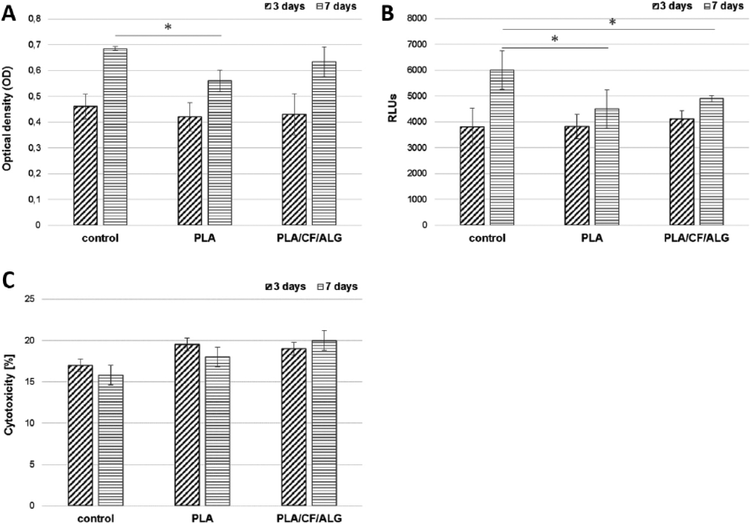
Cell viability, proliferation and cytotoxicity tests were conducted in quadruplicates at days 3 and 7. Cell viability was determined using the CellTiter assay (Promega, USA). Number (proliferation) of cells was assessed with a ToxiLight 100% Lysis test together with ToxiLight Bioassay kit (Lonza, USA). The intensity of bioluminescence obtained due to the reactions was related to the total adenylate kinase (AK) concentration released from all cells in the culture. Cytotoxicity of biomaterials was determined with the aid of a ToxiLight Bioassay (Lonza, USA) which measures AK released into the culture medium from damaged cells. All measurements were carried out with a microplate reader PolarStar Omega (BMG Labtech, Germany).
Results are reported as means ± standard deviation (SD). Statistical analyses were performed using the Student’s t-test (p<0.05).
Cells and their morphology were visualized with acridine orange (AO; Sigma, USA), a cell-permeable nucleic acid dye. AO can be used to detect apoptosis, as it shows chromatin condensation and formation of apoptotic bodies. Prior to observation, cells were stained for 1-2 minutes with 0.1% AO solution, rinsed with phosphate-buffered saline (Lonza, USA), and observed under a fluorescence microscope BX-41 (Olympus, Japan).
Animal implantation
The animal experiments were carried out on the basis of the approval of the Third Local Ethics Committee for Animal Experimentation in Warsaw, Poland. Animals were sedated using medetomidine (intramuscular injection; 0.4 mg/kg bodyweight [BWT]). Complex anesthesia was achieved with medetomidine (as above), then thiopental (intravenously; 50 mg/kg BWT) and isoflurane (inhalation; 1%-2.5% vol.). Euthanasia was performed with use of pentobarbital (intravenous injection; 150 mg/kg BWT).
Eight female Californian rabbits (approx. 9 months old) were used in the experiment. Five rabbits were implanted with prototype nails (diameter 2.5 mm, length 30 mm), and 3 rabbits were implanted with Kirschner wires (diameter 1.2 mm, length 30 mm) as a control. After reaching the distal end of femoral bone with a lateral parapatellar approach, perpendicular osteotomy was made with an oscillating saw 5 mm distal to the proximal end of the femoral trochlea. Next, bone fragments were fixed with 2 nails implanted through the articular surface, into predrilled channels. The ends of the nails were introduced as close to the articular surface as possible, shortened if needed. Appropriate bone fragment alignment and stability were achieved. The wound was closed in layers. After surgery, the rabbits were kept in cages. They were given antibiotics and analgetics for 3 postoperative days.
The evaluation was made on the basis of clinical observations, radiographs (anteroposterior [AP] and laterolateral [LL] projections) taken after 2, 4, 6 and 8 weeks post implantation, and macroscopic and histological observations. Histology samples were fixed in 10% buffered formalin, then decalcified in TBD-2 decalcifier (Shandon) and embedded in paraffin. Paraffin sections with a thickness of 4 µm were stained with hematoxylin and eosin (H&E) and evaluated under an optical microscope (Olympus BX 50).
Results
Cytotoxicity evaluation
The cytotoxic effects of PLA and PLA/CF/ALG were only slightly higher compared with the control (Fig. 1). Fluorescence microscopy observations (Fig. 2) confirmed the biocompatibility of the biomaterials studied, which was substantiated by a higher number of cells 7 days after seeding and good spreading of cells. AO-stained cells did not show any evidence of apoptosis (neither nuclei condensation nor apoptotic bodies were observed).
(
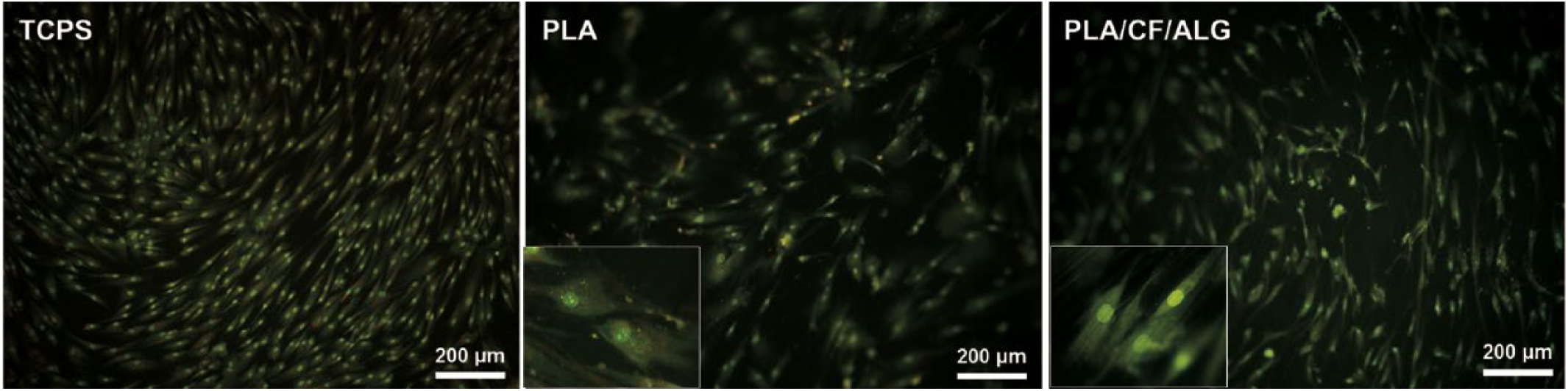
Fluorescence microscopy pictures of normal human osteoblast (NHOst) cell morphology after 7 days of culture on the surface of tissue culture polystyrene (TCPS) (
In vivo study
PLA/CF/ALG composite nails and control (Kirschner wires) were used for fixation of distal femoral osteotomy. One rabbit with composite nails was excluded due to humane endpoint before 2 weeks. Four rabbits implanted with composite nails used the limbs at 4 weeks post operation. In the control group, implanted with Kirschner wires, 2 rabbits loaded the limbs at 6 weeks, and 1 at 4 weeks.
Postsurgical radiographic images documented 1- to 2-mm fracture lines between bone fragments, which had sharp edges (Fig. 3); composite implants were radiolucent. At 2 weeks, the fracture lines were still clearly visible, but the edges had started to smooth out. At 4 weeks, shadows were visible in the fracture lines, as well as large amounts of additional shadows along the edges of fracture fragments. From 6 week onwards, the fracture lines became more radiopaque, signs of remodeling were present and the process of bony union had started. At 8 weeks, bony union and remodeling were recognized in all rabbits implanted with composite nails.
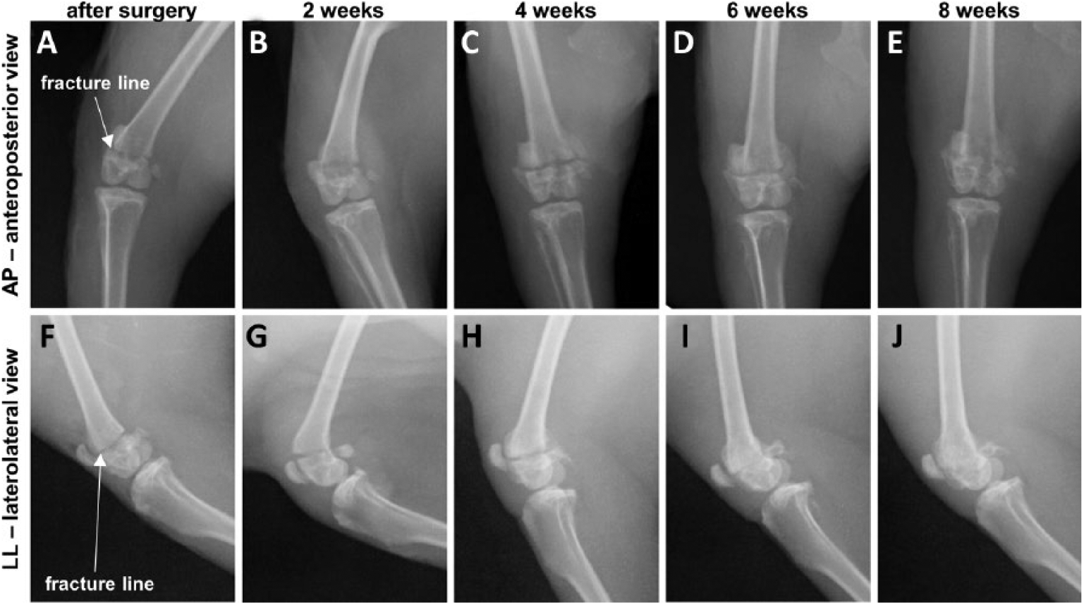
Radiographs of epiphysis fixation with polylactide/carbon fiber/alginate (PLA/CF/ALG) fiber nails: immediately after surgery – fracture lines with sharp edges visible between bone fragments (
In the control group (with Kirschner wires), radiographic images (Fig. 4) taken after surgery and at 2 weeks showed similar images of bone as described in the case of composite nails, but metallic wires used for fracture stabilization were visible. At week 4, in 1 rabbit, the fracture line had become wider; in another, shadows outside bony edges had appeared. After 2 more weeks, in 1 rabbit, the fracture line had narrowed, in another, bone union was recognized. In the third, bone remodeling and shadows outside bone edges were observed. At 8 weeks, bone union and bone remodeling were recognized in all rabbits implanted with Kirschner wires.
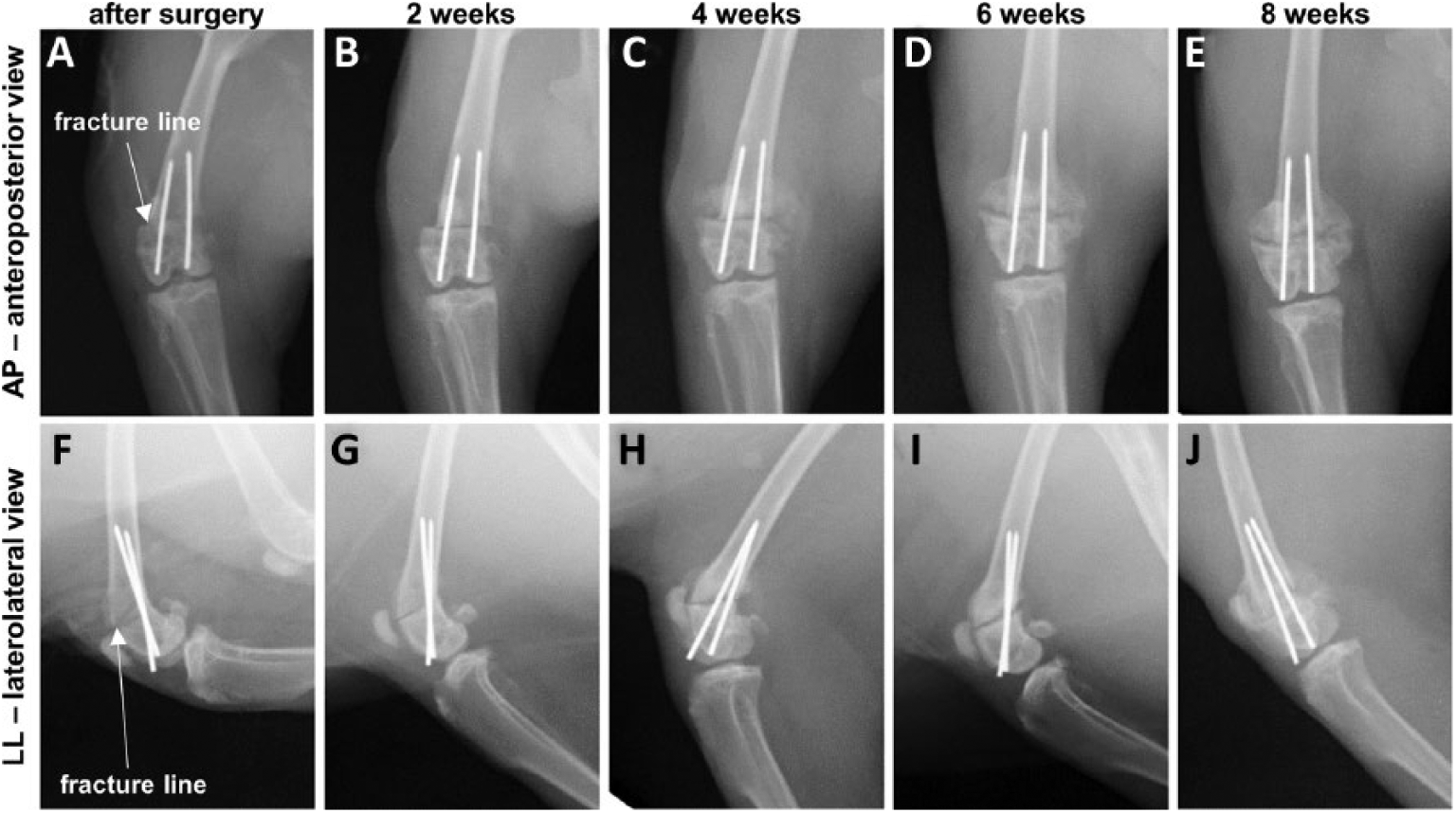
Radiographs of epiphysis fixation with Kirschner wires: immediately after surgery – fracture lines with sharp edges visible between bone fragments (
After the euthanasia of 1 rabbit at 4 weeks, distal ends of the PLA/CF/ALG nails were substantially covered with soft tissue, but bony union was not recognized. At 8 weeks, fractures had healed with large periosteal reactions. The end of 1 of the nails was covered with cartilage tissue. Composite nails, despite their consistency being soft, were hard to remove from the canals due to nail–tissue interaction and swelling of the ALG core and the whole nail. The diameter of the nail had increased by up to 4 mm (they had had a 2.5-mm diameter prior to implantation).
Post mortem macroscopic examination of osteotomies fixed with Kirschner wires performed 8 weeks after osteosynthesis (3 rabbits) revealed that the fractures were healed with large periosteal reactions. In 1 rabbit, 1 of the wires had become dislocated in such a way that it damaged the tibia and caused bleeding into the joint.
In the histological analysis at 4 weeks, fragments of carbon and ALG fibers in the form of conglomerates surrounded by granuloma were seen. Some signs of necrosis were also visible. After 8 weeks (Fig. 5), at the PLA/CF/ALG implantation site, loose connective tissue was present with both types of fibers from the slowly disintegrating implant. This was separated from the regular trabecular bone with fibrous connective tissue. Next to the remaining composite nail, numerous osteoblasts forming new bone trabeculae were seen.
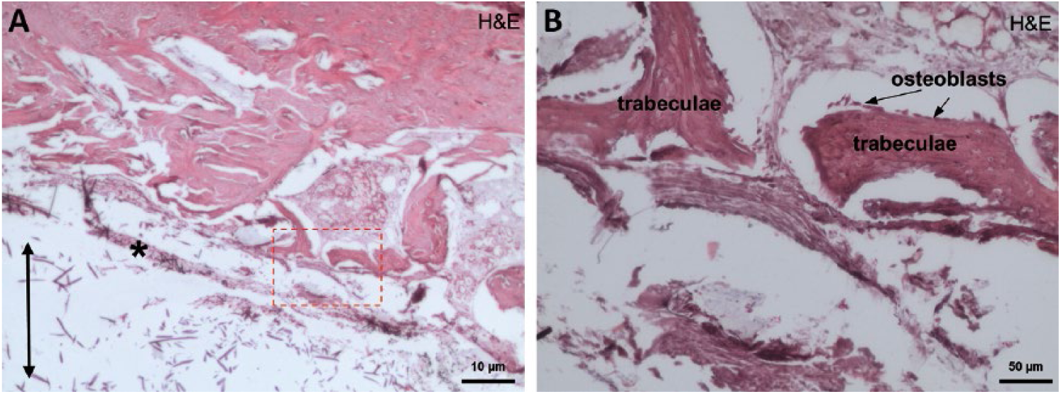
Histology showing tissue and polylactide/carbon fiber/alginate (PLA/CF/ALG) fiber nails used for fixation of distal epiphysis osteotomy after 8 weeks post implantation: (
Histological analysis of bone tissue contacting Kirschner wires at 8 weeks (Fig. 6), showed canals running from the articular surface into the bone, previously occupied by Kirschner wires removed before histopathological procedures. They contained necrotic masses, hyaline cartilage and bone remodeling foci located close to the canal.
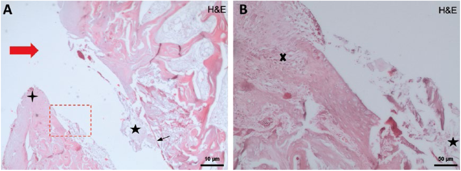
Histology showing tissues around Kirschner wire which were removed before histopathological preparation at 8 weeks: (
Discussion
The novelty of this work lies in its application of original, biodegradable composite intramedullary nails. The outer part of the nails consists of uniaxially oriented long carbon fibers, while the inner core is made of long ALG fibers. Both of the fibrous phases were saturated with PLA matrix.
Many studies have confirmed that PLA is a biocompatible material promoting cell proliferation and spreading, and that the addition of ALG enhances proliferation and differentiation of osteoblasts (13, 14). Furthermore, the addition of carbon fibers can trigger more rapid differentiation of osteoblasts (15). In our study, we observed only a small difference in viability and proliferation between samples of PLA and PLA/CF/Alg. Because of the presence of carbon fibers, processes of differentiation could predominate over those of proliferation (16, 17).
Cell tests in in vitro conditions as well as histopathology results confirmed the nails’ biocompatibility. There was no histological foreign body reaction visible. Osteoblast and osteoclast activity, important for bone healing and remodeling, were recognized on the basis of cell morphology due to destruction of epitopes during decalcification, which made more specialized staining impossible. Some particularly promising features of the PLA/CF/ALG nails were observed. The nails increased their volume after implantation into the bone. Moreover, their structure became softer with time, but the outer layer of carbon fibers anchored to the tissue. They were hard to remove from the bone, what can suggest that the nails may adjust to the bone canal.
Fixation of the osteotomy with the PLA/CF/ALG nails led to bony union. This was facilitated by large amounts of cancellous bone in the area and the introduction of 2 nails which gave stable fixation. Those were intra-articular fractures – in such cases, the presence of the synovial fluid hinders union. We speculate that the increase of the nail diameter resulted in sealing of the bone canal and together with strong anchorage of the fibers was responsible for a good result of osteosynthesis.
Histological analysis in the group implanted with the PLA/CF/ALG composite nails showed that the focal necrosis, observed especially at the beginning of the observation period, should be fully compensated by the osteoblast proliferation and trabeculae formation – signs of bone healing. At the same time, growth of cartilage at the ends of the nails just below the articular surface was observed macroscopically. The rabbits implanted with PLA/CF/ALG nails loaded the limbs early when compared with the control group implanted with Kirschner wires. This is another promising result; however, it should be noted that direct comparison to humans is not easy due to other relevant factors affecting recovery time that are hard to achieve in the case of animal studies. Those factors are type of rehabilitation and surgeon–patient cooperation, as well as the patient’s attitude and coexisting diseases. For example, in a series of cases of unicondylar fractures of distal femur in adult humans, rehabilitation therapy was started immediately in 65% of cases, with full weight bearing after 90 days (18).
Conclusion
In the current study, we were able not only do demonstrate the biocompatibility of our composite material in in vitro conditions but also the biocompatibility of the developed prototypes in in vivo conditions. We have also shown the potential applicability of PLA/CF/ALG nails for osteosynthesis of bone epiphysis. Further tests are necessary to reach the clinical study stage, but we strongly believe that the PLA/CF/ALG prototypes can be an alternative to metallic nails and can be translated into clinical practice.
Footnotes
Disclosures
Financial support: This work was supported by the Ministry of Science and Higher Education - Poland (research project no. 4575/B/T02/2009/37).
Conflict of interest: None of the authors has any financial interest related to this study to disclose.