
Abstract
Background
In this study, we investigated a method to produce bioactive hybrid amorphous silicon and silicon oxide patterns using nanosecond laser pulses.
Methods
Microscale line patterns were made by laser pulses on silicon wafers at different frequencies (25, 70 and 100 kHz), resulting in ablation patterns with frequency-dependent physical and chemical properties.
Results
Incubating the laser-treated silicon substrates with simulated body fluid demonstrated that the physicochemical properties of the laser-treated samples were stable under these conditions, and favored the deposition of bone-like apatite. More importantly, while NIH 3T3 fibroblasts did colonize the untreated regions of the silicon wafers, they showed a strong preference for the laser-treated regions, and further discriminated between substrates treated with different frequencies.
Conclusions
Taken together, these data suggest that laser materials processing of silicon-based devices is a promising avenue to pursue in the production of biosensors and other bionic devices.
Introduction
In bioengineering, there is a great demand for new materials to be used in biosensors, bionic devices, tissue engineering and cancer treatment (1, 2). Silicon is an attractive material for these mechanisms due to its semiconductor capabilities and mechanical properties. However, silicon is currently used primarily in microelectronics and photovoltaics (3–4–5–6–7–8). Its use in bionic devices is limited, as silicon is not biocompatible in its pure form (9, 10). The current solution is to package silicon in a bioactive material such as titanium (11). However, recent research demonstrates that a porous Si layer can be deposited onto silicon by chemical etching, improving biocompatibility and bioactivity (12). The chemical etching process is a lengthy procedure, has many complicated steps and results in the production of a great deal of waste. Also, the chemical etching process may introduce unknown toxins to a biological environment. Researchers have attempted to change the characteristics of silicon by using laser pulses, as laser ablation can also be used to generate a thin film of porous silicon on Si substrate (13, 14). In our previous studies, we found silicon nanoparticles on laser-treated Si surfaces, and showed hydroxyapatite (HA) deposition supporting its bioactivity (14).
Surface modification of biomaterials via laser ablation is becoming more popular. The 1,064-nm Nd:YAG laser, which is the same model of laser used in this study, has been used to oxidize the surface of titanium alloy implants. Radmanesh and Kiani found that the bioactivity of the implants was improved in vitro (15). Other lasers with varying wavelengths have successfully altered the surface of silicon-based materials (16, 17).
In this study, we introduced a unique method for fabrication of a hybrid amorphous silicon and SiO2 patterns on the silicon surface utilizing laser pulses, and showed that the laser-treated surfaces were preferential substrates for attachment and/or proliferation of mammalian fibroblast cells. These results have the potential to contribute to the development of cell growth manipulation technology in biosensors, bionic device fabrication and even cancer treatment (18).
Methods
Laser processing and generation of treated pattern
The laser used in this experiment was a nanosecond Nd:YAG pulsed laser manufactured by Bright Solutions. The laser has a wavelength of 1,064 nm, a maximum power output of 25 W, a maximum pulse energy of 1.5 mJ and a pulse duration of 8 ns. A simple line pattern was made on silicon wafers with orientation <100>. These patterns were made using EZCAD software and synthesized above the ablation threshold at a sub-microscale at different frequencies of 25, 70 and 100 kHz. The scanning speed of the laser was set to 100 mm/s, and the power was a mean of 15.1 W.
In vitro testing with simulated body fluid
Simulated body fluid (SBF) is a solution that approximates the ionic conditions of blood plasma, and is used to assess the bioactivity of a material by the evaluation of the growth of HA. Material that is able to have apatite form on its surface in SBF will have apatite produced on its surface inside the living body. This apatite layer has the ability to bond to living bone. This relationship holds as long as the material does not contain a component that induces toxic or antibody reactions. Examination of apatite formation on the surface of a material in SBF is useful for predicting the in vivo bioactivity of the material (19). One sample processed at a frequency of 100 kHz was incubated in SBF prepared according to the procedure specified by Kokubo and Takadama (19) at 36.5°C for 6 weeks.
Degradation testing with phosphate-buffered saline
Phosphate-buffered saline (PBS) is a fluid similar to SBF in that it also has similar ion concentrations to those of human blood plasma. This buffered solution was used to examine the spontaneous degradation of the treated silicon samples. Changes in sample ultrastructure were analyzed by scanning electron microscope (SEM) after incubation in PBS at 36.5°C for 6 weeks.
Culturing with NIH 3T3
We used National Institutes of Health 3T3 cell line (NIH 3T3) mouse embryonic fibroblast cells (American Type Culture Collection, Rockville, MD, USA) to characterize cellular interactions with the laser-treated Si samples. Triplicate cultures starting with 2 × 10⁵ cells were seeded in 60-mm dishes containing 1 sample of each variation of silicon substrate, and grown for 72 hours at 37°C under 5% CO2 in 4 mL Dulbecco's modified Eagle medium (DMEM), supplemented with 10% heat-inactivated calf serum, 4.5 mg/mL glucose and 2 mM glutamine. The silicon substrates were rinsed with PBS to remove nonadherent cells and fixed in 4% formaldehyde in PBS overnight at 4°C before staining and imaging.
Microscopy
Scanning electron microscopy
SEM was used to analyze samples, with a JEOL 6400 SEM equipped with Geller dPict digital image acquisition software and a Gatan ChromaCL Cathodoluminescence imaging system allowing capturing of high-resolution images.
Three-dimensional optical microscopy
We used a Zeta-20 Optical Profiler to obtain surface profiles.
Spectroscopy
Energy dispersive spectroscopy
To characterize elements present in the samples after laser treatment, we used energy dispersive spectroscopy (EDS). The model we used was a Hitachi SU-70 Field Emission Gun (FEG) SEM.
Micro-Raman spectroscopy
In addition to elemental analysis by EDS, we characterized chemical composition of the laser-treated samples using a Renishaw inVia micro-Raman spectrometer with a maximum power of 150 W.
Characterization of adherent cells
To characterize the distribution and morphology of the cells adhering to substrates, and their production of extracellular matrix (ECM) components, after fixation, cells were stained with Alexa Fluor 594-conjugated phalloidin (Thermo Fisher) to visualize the actin cytoskeleton and Draq5 as nuclear counterstain. Selected samples were also processed for indirect immunofluorescence using rabbit anti-fibronectin primary (Anaspec) and goat anti-rabbit Alexa Fluor 488 secondary (Thermo Fisher) antibodies (both diluted 1:1,000 in PBS + 0.1% Triton X-100 + 5% bovine serum albumin). Cells adhering to silicon substrates were imaged at low power using a Leica M205 stereo epifluorescence microscope to analyze the density of cells with respect to laser-treated regions of the silicon substrates, and at higher magnifications using a Leica SP-2 confocal microscope to analyze cytoskeletal architecture and characterize the production of fibronectin. Images were processed and analyzed using Fiji software (20).
Statistical Analysis
Statistical analysis must be utilized for this research for the surface topography of each silicon sample, as well as the line width and depth of the treated silicon surface. Proper statistical techniques help develop an efficient experimental design. All experiments were carried out in Minitab®. A minimum of 10 measurements were taken to obtain the mean height and width values of the sample profiles. Also, a p test was performed to compare adhesion preferences of each texture.
Results and discussion
To create a bioactive porous silicon layer, silicon nanofibers must be present on the silicon surface. In our previous study, these nanofibrous structures were observed on the surface of laser-treated silicon samples (14). In this work, we have extended this by characterizing how variations in laser treatments affect the ultrastructure and chemistry of silicon surfaces, and how these changes affect cellular interactions with these substrates. Figure 1 shows the SEM and 3D surface topography results for the treated silicon samples. It is clear that by changing the frequency, both width and depth of the laser-ablated area were considerably varied, which resulted in different surface roughnesses and surface energies. These changes in surface energy can significantly affect the cell adhesion of the irradiated areas.
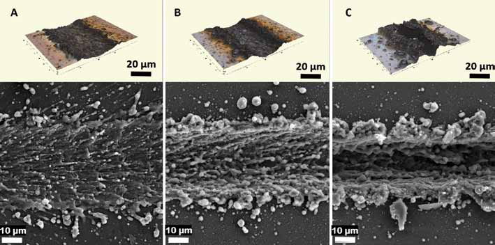
Three-dimensional optical microscopy images (upper) and scanning electron microscope (SEM) images (lower) of silicon samples, laser-treated at 25 kHz (
The cross-section of each silicon sample was plotted using 3D optical microscopy. This helped link the shape of the groove to the cellular response. These cross-sections are shown in Figure 2. The mean widths with respective standard deviations (±SD) of the grooves for 25 kHz, 70 kHz and 100 kHz were 45.8 ± 5.46 μm, 37.7 ± 5.41 μm and 19.7 ± 4.14 μm, respectively. However, as groove width decreased with increasing frequency, groove depth increased (Fig. 2). The groove depths, measured from the top-most peak to the bottom-most point of the trench, for 25 kHz, 70 kHz and 100 kHz were 4.8 ± 1.11 μm, 14.4 ± 0.92 μm and 17.1 ± 2.0 μm, respectively.
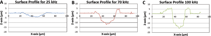
Cross-sections plotted using 3D optical microscopy to show textured areas for each frequency.
To evaluate the bioactivity of the fabricated structures, we used NIH 3T3 mouse embryonic fibroblast cells. Fibroblasts are the most common cell type in animal connective tissue. They play a critical role in normal wound healing which consists of closure of the wound, formation of granulation tissue and restoration of the vascular network and tissue architecture. Immigration of fibroblasts after injury, and their secretion and assembly of a functional ECM, lays the foundation for subsequent development of tissue architecture, including angiogenesis and the elaboration of more permanent connective tissues. The deposition and assembly of fibronectin by fibroblasts is widely regarded as the first crucial step in this process (21, 22).
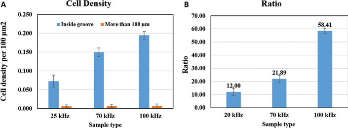
Interestingly, fibroblast cells showed a strong preference for the laser-textured regions of the samples (compared with the untreated regions) on samples treated with all 3 frequencies (Fig. 3). Further, there was a significant (p<0.01) difference between the densities of cells within the grooves generated by different laser frequencies, with the highest cell density observed at 100 kHz (Fig. 4). The effect of laser treatment on fibroblast colonization of the surface was significant, resulting in almost 60 times more cells per unit area associated with the 100-kHz textured surface than on the untreated silicon (Fig. 4).
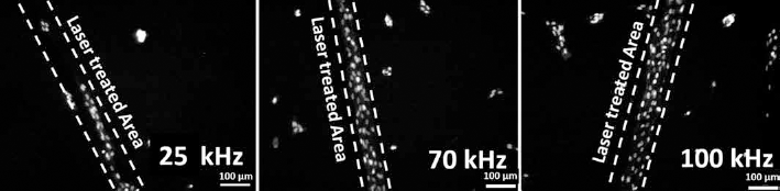
Epifluorescence microscopy images of Draq5 (nuclear)-stained cells adhering to samples treated with each frequency, showing increased cell density within treated areas.
(
Closer examination of the cells in the laser-etched grooves revealed that they possessed alignments in the textured path, especially with samples of a frequency of 100 kHz. Immunostaining also showed that they were secreting fibronectin (Fig. 5), an ECM protein secreted by fibroblasts during embryonic development and wound healing that lays the foundations for subsequent collagen deposition and tissue morphogenesis (22).
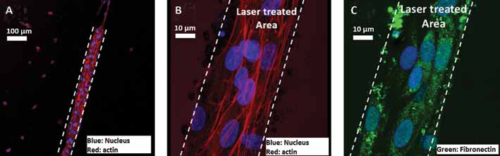
Confocal micrographs of fibroblasts in and around grooves etched at 100 kHz. Cells attached to untextured regions occurred at low density and had a rounded, unpolarized appearance (
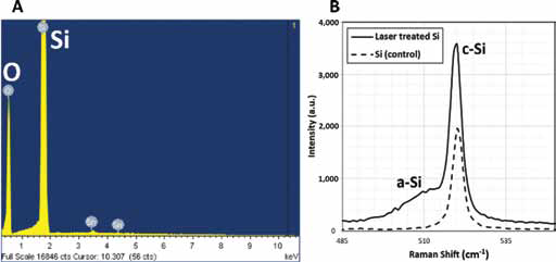
We used micro-Raman spectroscopy and EDS to identify the chemical composition of the silicon substrate (Fig. 6). The EDS results in Figure 6A show that the laser-treated silicon wafers consisted essentially of silicon and oxygen. The Raman results in Figure 6B show a sharp Raman shift at 520 cm−1 and a broad peak at 500 cm−1, confirming the presence of both crystalline silicon and amorphous silicon, respectively. A high-temperature reactive plume was generated at the contact area once the laser pulse irradiated the surface (23) (Fig. 7A). Some of the silicon ions reacted with the oxygen within and around the plume, creating silicon oxide, as shown in Figure 7B. The high thermal energy during the pulse excited the silicon ions so they assembled in a disordered pattern, which resulted in the formation of amorphous silicon, as illustrated in Figure 7C. The presence of amorphous silicon (a-Si) implies that during the cooling process, there was an undercooled liquid silicon layer. The cooling rate that is responsible for the formation of a-Si was too high to allow the nucleation of crystalline silicon (13). Once the pulse was terminated, the plume began to diminish in size and intensity. The lower thermal energy from the remaining plume allowed the silicon ions to organize into crystalline silicon, as shown in Figure 7D. At this point, the silicon ions had had time to cool to the crystalline phase. If the pulse separation time is decreased, or if the pulse frequency is increased, the amount of amorphous structures on the silicon surface after laser irradiation can be increased (18, 23). The correlation between the density of colonizing fibroblast cells and laser frequency suggests that the amount of hybrid SiO2 structure on the laser-treated surface may be related to the increase in bioactivity.
(
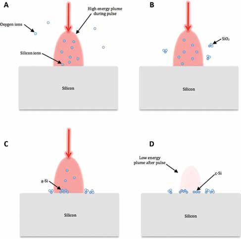
Plume generation and depletion with formation of amorphous silicon (a-Si), SiO2 and crystalline silicon (c-Si).
In vitro assessment using SBF is a method of evaluating the bioactivity of a material by testing the apatite-forming abilities of its surface (14, 23). We incubated 100-kHz samples in SBF for 6 weeks and assessed them using EDS (Fig. 8). We observed traces of sodium, chlorine, phosphorous and calcium present in the material after this treatment. These elements comprised bone-like apatite. The SiO2 in the substrate has a negative charge, which allows for calcium and phosphate ions to nucleate on the surface (24). This assembly of ions then allows for the formation of a bone-like apatite. This mechanism may explain the enhancement of bioactivity of the treated Si surface.
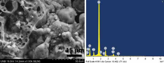
Scanning electron microscope (SEM) image of simulated body fluid (SBF) sample (left); and energy dispersive spectroscopy (EDS) results for the SBF sample (right).
To study the hybrid structure formation further, we evaluated the mean surface temperature during the laser treatment. To determine the mean surface temperature at the target area after a different number of laser pulses, we used theoretical methods from the relation between a laser pulse duration and absorbed intensity for optimum evaporation. From the heat conduction equation (Eq. [1]), we could determine the surface temperature after the end of a laser pulse (25, 26):
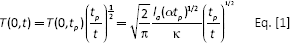
Where, tp is the pulse duration [s], Ia is the laser light intensity. The heat conduction coefficient and thermal diffusion coefficient in the case of silicon is 155 W/mK and 8.5 × 10⁻⁵ m2/s, respectively.
Directly before the laser ablates the surface, the surface temperature is at its minimum. When the laser hits the surface, the target area begins to absorb the energy for the duration of the pulse. At the end of the duration, the temperature of the surface is at its maximum, which can be written as Tmax or Tm = T(0,tp). The subsequent laser pulse is multiplied by the constant ratio for the previous maximum and the following minimum temperatures, α (Eq. [2]):
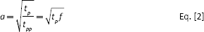
Where tp is the pulse duration in seconds, tpp is the pulse interval in seconds, which is equal to 1⁄f, and f is the pulse frequency in Hz.
Before and after each pulse, the maximum and minimum temperatures can be calculated as follows:
1st pulse:
2nd pulse:
nth pulse:
By using the absorbed laser light intensity Ia (Eq. [3]), the surface temperature can be simplified (Eq. [4]).
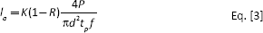
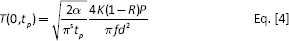
Where, K is the residual energy coefficient for silicon (0.8), R is the reflection coefficient, which varies with wavelength, P is the power in watts and d is the laser spot diameter in meters.
An interval equation to find the surface temperature at the ith laser pulse is as follows (Eq. [5]):
The average surface temperature after n pulses can then finally be calculated with Equation [6] (23, 24):
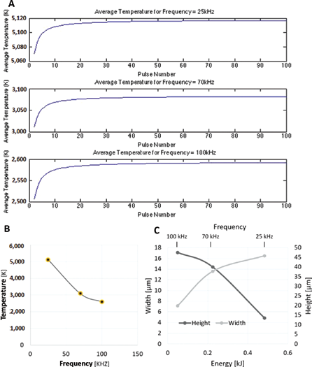
To simplify the temperature equations, some assumptions have been made. The assumptions in using these equations are that the silicon wafer samples are of adequate thickness, the heat-affected area is a point and there is no evaporation. Although evaporation would have an effect on the final value, these assumptions are made in order to obtain mean numbers for the purpose of studying the trend, not the actual surface temperature values (25). The mean surface temperature from Equation [6] was plotted against the number of pulses for each frequency, which can be seen in Figure 9A.
(
As seen in Figure 9A, the temperature settled at roughly 10 pulses for each frequency. These settling temperatures decreased with increasing frequency (Fig. 9B), as expected from the inverse correlation between pulse energy and frequency as per the performance plots supplied by the Nd:YAG laser manufacturer. At 25 kHz, 70 kHz and 100 kHz, the mean surface temperature was 3920 K, 2360 K and 1980 K, respectively, and the peak powers were 39.91 kW, 9.12 kW and 5.08 kW, respectively. The effective number of pulses for 25 kHz, 70 kHz and 100 kHz were 7.2, 20.2 and 28.8, respectively.
We then measured the height and width of each groove using a 3D optical microscope, and plotted the data against the pulse energy (Fig. 9C). As mentioned earlier, the depth of the treated area increased as the frequency was increased, and the groove got thinner (Fig. 2). The pulse energy change is an aspect of the Nd:YAG laser. As the frequency was increased, the pulse energy decreased, which resulted in a smaller width of a treated area, due to a smaller heat-affected zone. The higher frequency pulses had lower energy, resulting in lower average temperatures, but due to their more frequent passes, each pulse added to the previous pulse's existing trench, resulting in a deeper penetration. We also saw more of a wall built up along the sides of the groove in the 100-kHz sample compared with the 25-kHz sample (Figs. 1 and 2). This embankment of nanostructure may increase apparent cell adhesion by being able to trap cells within the hybrid structure-covered groove.
Nanostructured silicon can be degradable in vitro (27). Due to nanoparticles’ high degree of loading for drug molecules, the hybrid nanostructure may be useful for cancer treatment and drug delivery (28). For example, the degradability of these particles can reduce the side effects of chemotherapy. However, in the fabrication of biosensors and bionic devices, the degradation of nanostructured silicon may have adverse effects on an implant's structure. To investigate spontaneous degradation of the hybrid a-Si - SiO2 materials under physiological conditions, we conducted preliminary tests by incubating samples that varied in loop number in PBS. Loop number, or overlap number (OL), is the amount of times the laser overlaps the same pattern. The samples were made at 100 kHz, with loop numbers of 1, 3 or 5. Each sample was incubated in sterile PBS at 36.5°C for 4 weeks or 6 weeks, and the results are shown in Figure 10. The samples with varying frequency (Fig. 1) were made with 1 overlap.
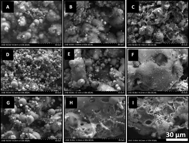
Phosphate-buffered saline (PBS) test: (
We saw no evidence of spontaneous degradation in the 1-OL samples (Fig. 10A–B–C); however, there did appear to be some degradation in the texture of the 3-OL (Fig. 10D–E) and 5-OL (Fig. 10G–H–I) samples during prolonged incubation in PBS. This raises the question of how laser-textured silicon materials may change over long periods in vivo, which will require further investigation.
Conclusion
Laser-treated silicon surfaces are more bioactive than untreated surfaces, as assessed by both their catalysis of the deposition of apatite from SBF and, more importantly, the response of fibroblast cells to treated vs. untreated areas of silicon substrates. These experiments do not allow us to distinguish mechanistically whether the increased cell density observed in the textured areas was due to preferential cell adhesion, increased cellular proliferation or decreased apoptosis (or some combination), but it is clear that the cells in the textured grooves are behaving more typically of cells in vivo. We are currently investigating the mechanism(s) underlying this observation.
SiO2 a-Si hybrid structure increases with frequency, and this correlates with increased cell density. Thus, it appears that the shape, phase and construction of the groove provided a favorable site for fibroblast cells. Given the facility with which modern materials science is able to manipulate surfaces using laser ablation, the possibilities for using this technology to manipulate cellular interactions with silicon structures are considerable.
Footnotes
Abbreviations
Financial support: This research is partially supported by grants from the New Brunswick Innovation Foundation (NBIF), the National Sciences and Engineering Research Council (NSERC) Discovery Grant program and the McCain Foundation to A.K., and by funding from the National Sciences and Engineering Research Council (NSERC) Discovery Grant program to B.D.C.
Conflict of interest: The authors declare that there is no conflict of interests regarding the publication of this paper.