
Abstract
Background
The aim of this study was to evaluate the effects of 3 different adhesives with different functional monomers, on the shear bond strength (SBS) of Biodentine®.
Methods
Acrylic blocks (n = 90) were prepared and a 2-mm height x 4-mm diameter hole was opened in each block. Every hole was completely restored with Biodentine®. Before preparation of composite restorations over the Biodentine® (2-mm height x 2-mm diameter), 3 different adhesives (Etch-37 (37%) w/BAC by Bisco & Prime Bond N&T, Clearfil S3 Bond and Adper Prompt L-Pop) were applied. SBS was evaluated using a universal testing machine, and failure mode for each sample was recorded. The results were statistically analyzed using 2-way ANOVA and post hoc Tukey test.
Results
When the megapascal values of all groups were compared, although there was no statistically significant difference in the different setting times (p>0.05), statistically significant differences were observed among all adhesive groups (p<0.05). Moreover, the highest SBS values were observed in the Clearfil S3 Bond group.
Conclusions
Clinical performance of Biodentine® may be affected by adhesive procedures and its setting time.
Introduction
Biodentine® has been introduced in recent years as a new tricalcium silicate-based form of restorative substance. Biodentine® powder consists predominantly of tricalcium silicate, which is supplemented with calcium carbonate and zirconium oxide. When combined with a water-reducing agent, the outcome is a calcium chloride solution that increases the rate at which the early strength of the bond develops and, therefore, results in a shorter setting time. As such, Biodentine® is preferred to mineral trioxide aggregate (MTA) because it offers greater viscosity and a reduced setting time of approximately 9 minutes (1).
Recent studies have indicated that Biodentine® provides a number of encouraging biological properties, especially in the areas of human pulp fibroblast cultures and on the dental pulp in an entire human tooth culture model (2, 3). One study, in which Biodentine® was applied directly to mechanically exposed rat pulps, revealed that this calcium-silicate based material has the ability to induce effective dentinal repair (4).
The esthetic qualities of resin composites mean that they are commonly used in the field of restorative dentistry. However, they cannot be applied directly on top of freshly mixed MTA since they have a negative impact on the rate and quality of the setting, and etching and rinsing unset MTA can dislodge the material. Biodentine®, with its reduced setting time of just 9 minutes, may represent a viable alternative to MTA because it is hypothetically possible for resin composites and glass ionomer cements (GICs) to be layered over set Biodentine® after 9 minutes, possibly allowing single-visit procedures (1, 5).
Self-etch adhesives are attractive to practitioners since they can be used without the requirement for a rinse phase. As such, their use can significantly reduce application time and technique sensitivity. In addition to their application after a 2-vs. a single-step approach, a further distinction can be made between “mild” and “strong” current self-etch adhesives (6, 7). Furthermore, the functional monomers that form an element of the self-etch adhesive offer polymerization qualities that bind the adhesive with the tooth structure or similar (8). Currently, very few studies (1, 5, 9) have examined the performance of Biodentine®, and as such, there is a lack of comprehensive knowledge and understanding of the strength with which restorative materials bond to Biodentine®. Previous studies evaluated performance of the clinical uses and bind to adhesives and restorative materials. In these studies, researchers randomly selected the adhesives (especially self-etch adhesives) (5, 9). The selection of adhesives according to ingredients was not addressed at all in this respect. In this study we focused on and selected the adhesives according to their pH values (≈0.8, ≈2.7 etc.) and functional monomers (dipentaerythritolpenta acrylate monophosphate [PENTA], hydroxyethylmethacrylate [HEMA], 10-methacryloyloxydecyl dihydrogen phosphate [10-MDP] etc.). The purpose of the current study was to assess the shear bond strength (SBS) of 3 different adhesive materials – an etch-and-rinse adhesive and 2 one-step self-etch adhesive materials – with different pH and functional monomer (HEMA, 10-MDP etc.), when used in combination with Biodentine®.
The null hypotheses tested were (i) Functional monomers will effect the bond strength test values and (ii) time intervals will have a positive effect on the SBS test.
Materials and method
Sample preparation
A schematic diagram representing the study sequence is given in Figure 1. The materials utilized in the study are listed in Table I. Acrylic blocks (n = 90) that contained a central hole with a height of 2 mm and a diameter of 4 mm were prepared. These holes were then completely filled with Biodentine® according to manufacturer instructions and smooth surfaces were obtained with amalgam condenser and burnisher ball (same as that used within an oral cavity) (Fig. 2). The specimens (n = 90) were subsequently stored at 37°C in 100% humidity. Half were set for 9 minutes, and half were set for 48 hours.
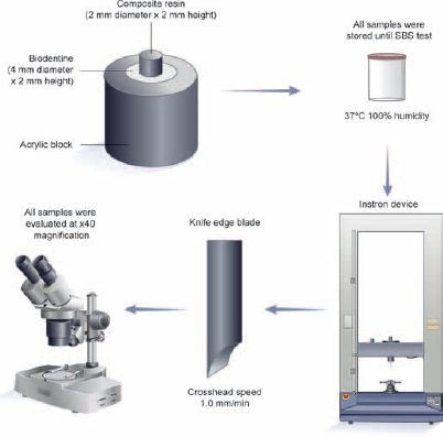
Schematic representation of the present study. SBS = shear bond strength.
Details of materials used in the study

BISEMA6 = bisphenol A polyethylene glycol diether dimethacrylate; Bis-GMA = bisphenol-A diglycidylemethacrylate; DMA = dimethacrylate; EDMAB = ethyl 4-dimethyl aminobenzoate; HEMA = hydroxyethylmethacrylate; 10-MDP = 10-methacryloyloxydecyl dihydrogen phosphate; TEGDMA = triethylene glycol dimethacrylate; UDMA = diurethane dimethacrylate.
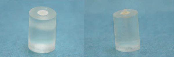
Prepared sample.
After the setting time had elapsed, the samples were randomly divided into 3 groups of 30 specimens each, as follows:
Group 1: Clearfil S3 Bond (Kuraray Medical, Osaka, Japan) (9 minutes, 48 hours);
Group 2: Adper Prompt L-Pop (3M/ESPE, St. Paul, MN, USA) (9 minutes, 48 hours);
Group 3: Total Etch System (Acid: Etch-37 (37%) w/BAC; Bisco, USA) in conjunction with Bond (Prime Bond N&T, Dentsply, Germany) (9 minutes, 48 hours).
Materials used in this study were applied according to the manufacturer's instructions (Tab. I).
Following preparation, the Biodentine® material was placed in cylindrical plastic tubes of 2-mm height and 4-mm diameter, so that each resin composite specimen was placed at the center of the Biodentine® surface. The composite (Filtek Z250, 3M/ESPE, St. Paul, MN, USA) specimens were then cured for 20 seconds using a light-emitting diode light cure (VALO LED; Ultradent, South Jordan, UT, USA) at an intensity of 1,200 mW/cm2. Following the polymerization, or setting procedure, the plastic tubes were carefully removed from the specimens and stored at 37°C in 100% humidity for 48 hours. To ensure consistency, a single technician prepared and tested all samples.
SBS test preparation
The mounted samples were subjected to a SBS test in a universal testing machine (Instron; Shimadzu Corp., Tokyo, Japan). A chisel-edge plunger (knife-edge blade) was mounted on the movable crosshead (crosshead speed: 1.0 mm/min) of the test machine and positioned in such a manner that the leading edge was targeted at the Biodentine® base/adhesive interface. The total force required to remove the restorative material was measured in Newtons (N), and the SBS was then calculated by dividing the peak load values by the restorative material base area (πr2 = 3.14 mm2).
Fracture analysis
The fractured samples were stored in distilled water for a period of 24 hours following the SBS testing procedure. Failure modes were evaluated at ×40 magnification with a stereoscopic microscope. The failure mode for each sample was then assessed according to one of the following classifications (10):
Adhesive failure: The failure was at the interface between restorative material and Biodentine® (bonding area).
Mixed failure: The failure was a combination of interfacial separation and partial cohesive failure of the Biodentine® and/or restorative material.
Cohesive failure: The failure was a fracture within the Biodentine® or restorative material.
Statistical Analysis
All data were entered into statistical computer software (SPSS ver. 20; SPSS Inc., Chicago, IL, USA). Two-way ANOVA and post hoc Tukey tests were used. All data were tested at a significance level of 0.05.
Results
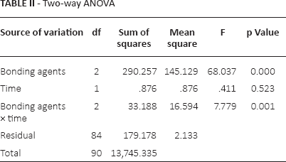
The statistical analysis is shown Table II and Figure 3. Overall, all adhesive groups were evaluated; there was no statistically significant difference between the 9-minute group and the 48-hour group 48-hour in each adhesive material (p>0.05).
Two-way ANOVA
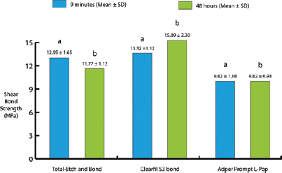
Mean of shear bond strength values (MPa) of 3 adhesive systems. There was no statistically significant difference between columns with the same letters.
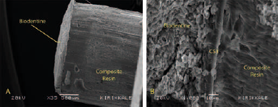
After the 9-minute period, whereas there was no statistically significant difference between Total Etch and Clearfil S3 Bond (p>0.05), a statistically significant difference was observed between Total Etch and Adper Prompt L-Pop (p<0.05). Also, a statistically significant difference (p<0.05) between SBS values for Clearfil S3 Bond and Adper Prompt L-Pop was observed. There were statistically significant differences among all adhesive groups, after the 48-hour period (p<0.05). Adper Prompt L-Pop showed the lowest SBS after the 9-minute and 48-hour periods. The highest SBS values were observed in Clearfil S3 Bond. Figure 4 summarizes the failure patterns of the specimens at 9-minute and 48-hour intervals. Cohesive fracture was detected as the most common fracture type. A scanning electron microscopy (SEM) image of fractured Biodentine® with the Clearfil S3 Bond sample, demonstrating cohesive failure, is shown in Figure 5, and SEM images of Biodentine® material after SBS test is shown at different magnifications (×35, ×350, ×1,000 and ×4,000) in Figure 6 (cohesive failure), Figure 7 (mixed failure) and Figure 8 (adhesive failure).
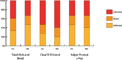
Fracture types of tested materials.
(
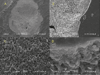
Scanning electron microscopy (SEM) image of the Cohesive failure of Biodentine® material after shear bond strength (SBS) test, shown at ×35
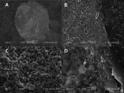
Scanning electron microscopy (SEM) image of the mixed failure of Biodentine® material after shear bond strength (SBS) test, shown at ×35
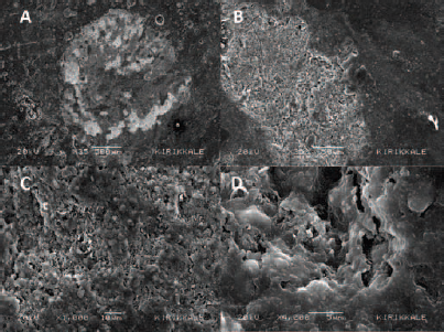
Scanning electron microscopy (SEM) image of the adhesive failure of Biodentine® material after shear bond strength (SBS) test, shown at ×35
Discussion
Biodentine® may be a successful material, when it is used instead of MTA. Biodentine® has a shorter setting time, good placement and bioactivity, compared with MTA (1, 11). In the literature, there is little information about SBS testing of Biodentine® combined with different adhesives and restorative materials (5, 9, 12). The aim of the present study was to evaluate the SBS performance of Biodentine® using 3 different adhesives, which have different functional monomer and pH values, in terms of 2 time intervals.
The first null hypothesis was accepted because statistically significant differences were observed among all adhesive groups. The second null hypothesis was rejected. When compared with each other, there were no statistically significant differences between 9-minute and 48-hour time intervals in all groups.
Although many functional monomers have been incorporated in different adhesives for improving bonding abilities to teeth, some researchers have reported that the functional monomer 10-MDP may chemically bind to calcium ions in Biodentine®, and because of this, it improves micromechanical attachment and chemical adhesion between them (8, 12). In the present study, we used the Clearfil S3 Bond, which includes the 10-MDP functional monomer; we thought that it could increase the bond strength performance of Biodentine®. According to the findings of the present study, Clearfil S3 Bond groups showed the highest SBS values, which is line with previous research into functional monomers (8, 12).
In the present study, there was no statistically significant difference among all groups in terms of time intervals (p>0.05), but SBS values decreased over time, except in the Adper Prompt L-Pop bond groups. In clinical practice, Biodentine® may be more effective and successful, if it is used as an underlying material for restorative materials. When Biodentine® materials are used with bonding agents, which include 10-MDP functional monomer, the binding ability of Biodentine® to restorative dental materials will increase.
While the Clearfil S3 Bond groups showed the highest SBS values, the Adper Prompt L-Pop bond groups showed the lowest values in present study. SBS values of the Total Etch group were similar to those of the Clearfil S3 Bond group. However, a statistically significant difference was observed between these groups after 48 hours. Odabaş et al (9) reported that Clearfil SE Bond showed the highest values (at 12 minutes, 16.90 ± 8.11 MPa; at 24 hours, 19.56 ± 7.58 MPa) and Clearfil S3 Bond values were the second highest (at 12 minutes, 11.06 ± 3.85 MPa; at 24 hours, 15.19 ± 3.34 MPa). Although, SBS values recorded for the Clearfil S3 Bond from this study were found to be consistent with the results of the present study (Clearfil S3 Bond at 10 minutes was 13.32 ± 1.12 MPa, at 48 hours, 15.09 ± 2.30 MPa), our results were higher than their results (9). These differences may be explained by different operators and time intervals.
Tay and Pashley (7) reported that self-etch adhesives have different degrees of aggressiveness. Therefore, researchers have classified self-etch adhesives according to their pH values; mild self-etch adhesive (pH >2), moderate self-etch adhesive (1< pH <2) and aggressive self-etch adhesive (pH <1) (6, 13). Aggressive self-etch adhesives have deep demineralization effects on the dentin and dentin-like materials, because of their high acidities. On the other hand, mild and moderate adhesive systems have only up to 1-μm depth of demineralized dentin (14). In the present study, 2 different adhesives were used, one of them aggressive and the other mild. Their pHs were as follows: Adper Prompt L-Pop pH ≈0.8, Clearfil S3 Bond pH ≈2.7.
According to our results, the mild adhesive agent (Clearfil S3 Bond) bonded to Biodentine® more effectively than the aggressive adhesive agent (Adper Prompt L-Pop). This difference may be explained by the presence of the functional monomer 10-MDP in Clearfil S3 Bond.
It must be emphasized that the results of this study cannot be directly extrapolated to in vivo situations. The present study was a laboratory investigation, and the experiment was performed at room temperature. Different results may have been achieved with intraoral conditions.
Forthcoming studies should examine the effects of different bonding procedures on the surface of primary and permanent tooth, compared with Biodentine®. To increase our knowledge about Biodentine®, many in vitro studies need to be performed with different laboratory procedures and conditions.
Conclusions
Within the limitations of this in vitro study,
Biodentine® materials may be clinically more effective and useful, if they are used with self-etch adhesive systems. Especially, Clearfil S3 Bond, which contains 10-MDP, may be more effective.
Biodentine® may be a good alternative to MTA, because of its quick setting and application time.
Footnotes
Financial support: No grants or funding have been received for this study.
Conflict of interest: None of the authors has financial interest related to this study to disclose.