
Abstract
Background
Cu-Al-Ni shape memory alloys (SMAs) possess two-way shape memory effects, superelasticity, and damping capacity. Nonetheless, Cu-Al-Ni SMAs remain promising candidates for use in biomedical applications, as they are more economical and machinable than other SMAs. Ensuring the biocompatibility of Cu-Al-Ni SMAs is crucial to their development for biomedical applications. Therefore, this study aimed to assess the toxicity of Cu-Al-Ni SMAs using a Probit dose–response model and augmented simplex design.
Methods
In this study, the effects of Cu2+, Al3+ and Ni2+ metal ions on bacteria (Escherichia coli DH5α) using Probit dose–response analysis and augmented simplex design to assess the actual toxicity of the Cu-Al-Ni SMAs.
Results
Extraction and repetition of Escherichia coli DH5α solutions with high Cu2+ ion concentrations and 30-hour incubation demonstrated that Escherichia coli DH5α was able to alter its growth mechanisms in response to toxins. Metal ions leached from Cu-Al-Ni SMAs appeared in a multitude of compositions with varying degrees of toxicity, and those appearing close to a saddle region identified in the contour plot of the augmented simplex model were identified as candidates for elevated toxicity levels. When the Cu-13.5Al-4Ni SMA plate was immersed in Ringer's solution, the selective leaching rate of Ni2+ ions far exceeded that of Cu2+ and Al3+. The number of Cu2+, Al3+ and Ni2+ ions leached from Cu-Al-Ni SMAs increased with immersion time; however, at higher ratios, toxicity interactions among the metal ions had the effect of gradually reducing overall toxicity levels with regard to Escherichia coli DH5α.
Conclusions
The quantities of Cu2+, Al3+ and Ni2+ ions leached from the Cu-13.5Al-4Ni SMA plate increased with immersion time, the toxicity interactions associated with these compositions reduced the actual toxicity to Escherichia coli DH5α.
Introduction
Fe-Cr-Ni stainless steels, Co-Cr–based alloys, pure Ti metal and Ti-6Al-4V alloys possess the biocompatibility needed for biomedical applications (1). These materials are typically used in implants for medical and dental applications, joint replacement, fixation of bone fractures and orthopedic purposes (2-3-4). Shape memory alloys (SMAs) are another advanced biomaterial with unique shape memory effect and superelasticity (5). Among these, nickel-titanium (TiNi) SMAs are the most widely investigated due to their favorable biofunctionality in specific biomedical applications, such as laparoscopic surgery, intracoronary stents and ligament replacement, and for bone stamp and osteosynthesis devices, as well as in shape memory microvalves for the control of drug delivery (6-7-8). Conversely, Cu-Al-Ni SMAs possess 2-way shape memory effects, superelasticity and damping capacity. However, they also exhibit a recoverable nonlinear superelastic strain of approximately 18%, greater than that of normal TiNi SMAs (9-10-11-12-13). Nonetheless, Cu-Al-Ni SMAs remain promising candidates for use in biomedical applications, as they are more economical and machinable than other SMAs (14-15-16). Čolić et al (17) previously performed an in vitro investigation of the microstructure, corrosion behavior and cytotoxicity of rapidly solidified Cu-Al-Ni SMA ribbons. These researchers reported that rapid solidification significantly improved corrosion resistance and biocompatibility. Nevertheless, any degradation in the biocompatibility of Cu-Al-Ni SMA implants poses a risk of metal ions being released (18). Ensuring the biocompatibility of these materials is crucial to their development for biomedical applications; therefore, this study investigated the concentrations of the Cu2+, Al3+ and Ni2+ metal ions in Ringer's solution following selective leaching from Cu-Al-Ni SMAs. We further examined the effects of Cu2+, Al3+ and Ni2+ metal ions on bacteria (Escherichia coli DH5α) using Probit dose–response analysis and augmented simplex design (19) to assess the actual toxicity of the Cu-Al-Ni SMAs.
Methods and materials
Probit dose–response model
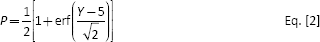
The dose–response curves of Cu, Al and Ni metal ions were analyzed using a Probit dose–response model, in which the tolerance of bacteria to toxic compounds in a given population exhibited a log-normal distribution (20). On the dose-response curve, the half-maximal effective concentration (EC50) is typically used to compare toxicities of metals because it is simpler to apply and can be more accurately interpolated than EC0 and EC100, which were normally extrapolated estimated. The semi-logarithmic plots of metal ion concentrations are assumed to behave in a linear relationship with the obtained responses. Thus, the Probit dose–response model converts a sigmoidal-shaped dose–response curve into a linear normal equivalent deviation (NED) scale. A Probit unit (P) in the model is equal to a NED scale value plus 5. The formulae for determining concentrations are as follows (20-21-22-23):

where A and B are the intercept and Hill slope of the dose–response relationship; Z is the concentration of metal ions (mg/L); Y is the Probit unit; P is the response (%) corresponding to the target metal; and erf (x) is an error function. Note that the response variable is normalized to fall between 0 and 1. The conversion relationship for provoked responses has previously been outlined by Chen et al (21, 22). Response P can be determined as follows:
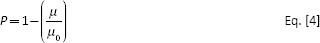
where μ denotes the maximum specific growth rate. Kinetic parameter μ is determined according to the specific growth curves of the cultures in the experiment. Microbes grown in a metal-laden environment often demonstrate decreased growth rates. Therefore, the reference parameters μ0 are determined from growth curves corresponding to various concentrations of target metals, rather than a concentration of zero in a metal-free culture (21). All inocula used in this study were selected from precultures performed in metal-free media.
Incubation of E. coli DH5α
The toxicity assessment of the Cu-Al-Ni SMAs was established by the growth of E. coli DH5α using Probit dose–response analysis and augmented simplex design. We selected E. coli DH5α as a study species due to their easy incubation and sensitivity to toxic elements. The Cu, Al and Ni metal ions used in Probit dose–response analysis and augmented simplex design originated from high-purity atomic absorption spectroscopy (AAS) standard solution (1,000 mg/L) purchased from Merck. Each AAS solution was diluted to various concentrations, and mixed with sodium citrate buffer solution (0.5 × Luria-Bertani [LB]) and deionized water to formulate metal ion solutions ranging between 0 and 1,000 mg/L. E. coli DH5α was precultured in LB broth (10 g/L tryptone, 5 g/L yeast extract, 10 g/L NaCl) at 37°C, under stirring at 125 rpm for 12 hours. The cultured E. coli DH5α solutions were then added to each solution of metal ions at a volume ratio of 1:100. The concentration of E. coli DH5α was determined by measuring the optical density (OD) value of the cell solution every 30 minutes for the initial 5 hours, and then every 1 hour up until 24 hours. The OD values were measured at 600 nm (OD600) using a spectrophotometer (GENESYS 20; Thermo Scientific) with cultured broth as the blank cuvette.
Selective leaching of Cu-13.5Al-4Ni SMA
A plate of Cu-13.5Al-4Ni SMA was used for selective leaching tests due to the pronounced martensitic transformation and shape memory properties of this chemical composition (13). The Cu-13.5Al-4Ni plate was prepared from pure raw copper (purity 99.9 wt%), aluminum (purity 99.99 wt%) and nickel (purity 99.9 wt%). The raw materials were melted at 1100°C in evacuated quartz tubes followed by quenching in ice water. The quenched ingots were annealed at 900°C for 30 minutes and then allowed to cool slowly in the furnace until reaching room temperature. The annealed Cu-13.5Al-4Ni SMA ingots were cut into plates with dimensions of 30.0 × 6.5 × 1.3 mm3 for the selective leaching test. The Cu-13.5Al-4Ni SMA plates were then immersed in a test flask containing 250 mL of Ringer's solution and maintained at 37°C in an orbital shaker incubator for 60 days to determine their selective leaching behavior. The precise concentrations of Cu, Al and Ni ions in the Ringer's solution were determined using inductively coupled plasma-mass spectrometry (ICP-MS, Agilent 7500ce) following the leaching process.
Results and discussion
Growth behaviors of bacteria cultured with metal ions
Figure 1A and B presents the lnOD600 = Natural logarithm of the optical density measured at 600 nm values of E. coli DH5α solutions combined with Al3+ and Ni2+ ion solutions of various concentrations as a function of time. The lnOD600 value of E. coli DH5α without the addition of any metal ions (control group) is also plotted for comparison. Figure 1A and B shows that the lnOD600 values for each curve were extremely small in the first hour. The delayed growth of E. coli DH5α in this stage is referred to as the lag time. Between 1 hour and 6 hours, the lnOD600 values increased noticeably, indicating the exponential growth stage of the E. coli DH5α. Following this exponential growth stage, the lnOD600 values for each curve gradually leveled off until reaching a static state at 24 hours. Figure 1 also reveals that increasing the concentration of Al3+ and Ni2+ ions led to lower lnOD600 values for E. coli DH5α. Taken together, our results demonstrate that the addition of Al3+ and Ni2+ ions in solution can inhibit the growth of this bacterial strain.
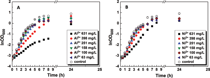
The optical density (lnOD600) = Natural logarithm of the optical density measured at 600 nm values of Escherichia coli DH5α solutions combined with Al3+ (
Figure 2A presents the lnOD600 values as a function of time for E. coli DH5α cultured with Cu2+ ion solutions of various concentrations. Unlike Al3+ and Ni2+ ions, higher concentrations of Cu2+ ions increased the lag time of E. coli DH5α; lag tine reached 10 hours when the concentration of Cu2+ ion was 631 mg/L. This indicates that the growth of E. coli DH5α can be impeded by a sufficient concentration of Cu2+ ions. To further elucidate the toxic effects of these ions, the E. coli DH5α solutions with high Cu2+ ion concentrations (398, 500 and 631 mg/L) and 30-hour incubation were extracted and subjected to the identical tests. The results are shown in Figure 2B. Figure 2B shows that the lag time of the E. coli DH5α solutions was shortened considerably under these treatment conditions, entering a phase of rapid growth after 1 hour. These results demonstrate that E. coli DH5α is able to alter its growth mechanisms in response to toxins, such as high concentrations of Cu2+ ions.
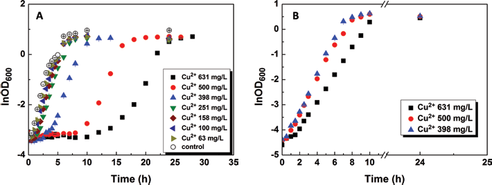
(
Tolerance of bacteria to metal ions
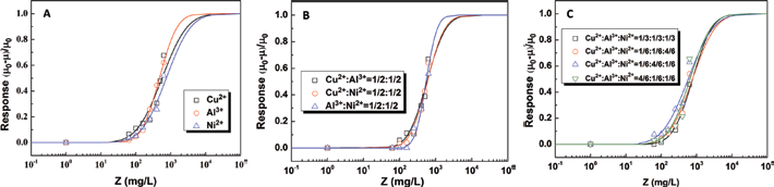
Figure 3A presents the dose–response curve plotted from the relationship between response P, and concentration Z for single Cu2+, Al3+ and Ni2+ ions. From Figure 3A, the EC50 values for Cu2+, Al3+ and Ni2+ ions were calculated as 563.5, 474.1 and 745.1 mg/L, respectively. This indicates that the relative toxicities of these metal ions with respect to E. coli DH5α were as follows: Al3+ > Cu2+ > Ni2+. Figure 3B and 3C plots the dose–response curves for binary and ternary metal ions, respectively, under various concentration ratios. From Figure 3B, the EC50 values were as follows: Cu2+: Al3+ = 1: 1 (572.9 mg/L), Al3+: Ni2+ = 1: 1 (544.5 mg/L) and Cu2+: Ni2+ = 1: 1 (588.8 mg/L). Thus, the toxicity of binary ions at ratios of Cu2+: Al3+ = 1: 1 and Cu2+: Ni2+ = 1: 1 are comparable and slightly lower than at a ratio of Al3+: Ni2+ = 1: 1. These results demonstrate that toxicity interactions between metal ions may cause synergism, antagonism or additive effects in binary metal ions systems. The EC50 values in Figure 3C were as follows: Cu2+: Al3+: Ni2+ = 1: 1: 1 (768.4 mg/L), Cu2+: Al3+: Ni2+ = 1: 1: 4 (762.4 mg/L), Cu2+: Al3+: Ni2+ = 1: 4: 1 (578.0 mg/L) and Cu2+: Al3+: Ni2+ = 4: 1: 1 (648.2 mg/L). The fact that the toxicity of Cu2+: Al3+: Ni2+ = 1: 4: 1 slightly exceeded that of other concentration ratios can be attributed to the higher toxicity of Al3+ ions. However, this is not an issue of concern, due to the complex interactions between these 3 metal ions and subsequent effects on toxicity. Hornez et al (24) have reported that aluminum has excellent cytocompatibility; however, there is a chance that a small quantity of Al ions released from Cu-Al-Ni SMAs may modify the activities of Cu and Ni ions.
Dose–response curves of Cu-Al-Ni single (
Assessment of toxicity in the Cu-Al-Ni ternary system
The toxicity of the Cu-Al-Ni ternary system on E. coli DH5α was further assessed using an augmented simplex design, which is the most common approach to dealing with mixtures. As shown in Figure 4, the factorial space comprising all possible compositions of the constituent components in a Cu-Al-Ni ternary system forms a triangle, of which the vertices correspond to ions of Cu2+ (point A), Al3+ (point B) and Ni2+ (point C). In addition to the vertices, the central point of the triangle (point G), the midpoints along the lines connecting the vertices (points D-F) and the mid-points between the central point and vertices (points H-J) are also considered in the augmented simplex design (25). All responses from points A to J can be formulated using a typical quadratic model as follows:
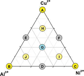
Schematic diagram of triangle ternary graph for Cu-Al-Ni augmented mixture design.

where x, y and z represent the concentration ratios of Cu2+, Al3+ and Ni2+, respectively. Figure 5A and B respectively represents a 3-dimensional plot and a 2-dimensional contour plot of the formulated quadratic model. As shown in Figure 5B, the EC50 values present a hump denoted as region I, and 2 saddles denoted as region II. Accordingly, we can assume that the toxicity of the implant material is likely to be higher in cases where the chemical composition of metal ions leaching from implant material in the vicinity of region II. Conversely, the biocompatibility of materials used for implants is likely to be higher in cases where the chemical composition of the leached metal ions is close to region I. The hump region I shown in Figure 5B indicates that the nickel ion is relatively biocompatible in this study. This feature matches with the results that the incubation of E. coli DH5α cultured with Ni2+ ion solutions was inhibited during the lag phase, but the difference seems to disappear with longer incubation, as demonstrated in Figure 2. Therefore, toxicity of Cu-Al-Ni SMAs with various chemical compositions can be preliminarily predicted and verified efficiently using E. coli DH5α. However, further investigations are still required to determine the actual toxicity of metal ions released from implant materials into surrounding tissues.
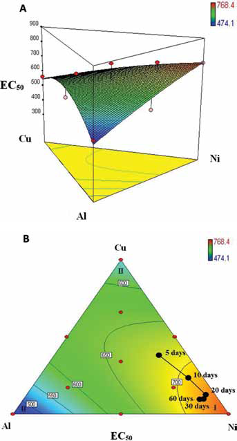
(
Selective leaching of Cu-Al-Ni SMAs
Figure 6 presents the concentrations of Cu2+, Al3+, and Ni2+ ions following selective leaching from a plate of Cu-13.5Al-4Ni SMA immersed in Ringer's solution. As shown in Figure 6, the concentration of Al3+ ions was extremely low (below 60 ppb), ppb = parts per billion even after immersion for 60 days. Cu2+ ions presented a higher concentration of approximately 170 ppb after immersion for 10 days, and remained at that level for the duration of the 60-day study period. At the same immersion time, the concentration of Ni2+ ions far exceeded those of Al3+ and Cu2+ ions, and approaching 850 ppb after 60 days. Clearly, the Ni2+ ions leached from the Cu-13.5Al-4Ni SMA plate far more rapidly than the Al3+ and Cu2+ ions. Here, it should be noted that the leaching characteristics depend on media and condition used to perform the experiment. In Figure 5B, the composition ratios of the metal ions selectively leached from the Cu-13.5Al-4Ni SMA plate are plotted according to immersion time. This plot illustrates that the composition of metal ions gradually approached hump region I as immersion time increased. These findings suggest that despite an increase in Cu2+, Al3+ and Ni2+ ions leached from the Cu-13.5Al-4Ni SMA plate with immersion time, the actual toxicity to E. coli DH5α was reduced as a result of stringer toxicity interactions between ions. Therefore, Cu-13.5Al-4Ni SMA should be biocompatible for use in biomedical applications. However, the relatively high concentrations of leached Ni2+ ion may pose a risk in biomedical applications, further appropriate surface modification is suggested if Cu-Al-Ni SMAs are to be used as implant materials.
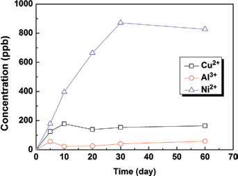
Concentrations of Cu2+, Al3+ and Ni2+ ions following selective leaching from a plate of Cu-13.5Al-4Ni SMA immersed in Ringer's solution. ppb = parts per billion.
Conclusions
This study sought to determine the toxicity of a 3-component Cu-Al-Ni SMA with an arbitrary chemical composition, using the Probit dose–response model and the augmented simplex method. Al3+ and Ni2+ ions are known to inhibit the growth of E. coli DH5α. Nonetheless, the growth mechanism of E. coli DH5α was able to adapt to a toxic environment containing high concentrations of Cu2+ ions. A contour plot of the EC50 values for Cu-Al-Ni SMAs presents a hump near pure Ni2+ and 2 saddles close to pure Al3+ and Cu2+. A composition of metal ions leached from Cu-Al-Ni SMAs located close to the saddle region, could be an indication of elevated toxicity levels. Additionally, when a Cu-13.5Al-4Ni SMA plate was immersed in the Ringer's solution, the selective leaching rate of Ni2+ ions was far higher than that of Cu2+ and Al3+. Finally, despite the fact that the quantities of Cu2+, Al3+ and Ni2+ ions leached from the Cu-13.5Al-4Ni SMA plate increased with immersion time, the toxicity interactions associated with these compositions reduced the actual toxicity to E. coli DH5α.
Footnotes
Financial support: The authors gratefully acknowledge the financial support for this research provided by the National Science Council (NSC), Taiwan, Republic of China, under Grant No. NSC102-2221-E-197-006.
Conflict of interest: The authors declare that they have no conflict of interest.