
Abstract
Carbon nanotubes (CNTs) have been widely recognized and used for controlled drug delivery and in various other fields due to their unique properties and distinct advantages. Both single-walled carbon nanotubes (SWCNTs) and multiwalled (MWCNTs) carbon nanotubes are used and/or studied for potential applications in medical, energy, textile, composite, and other areas. Since CNTs are chemically inert and are insoluble in water or other organic solvents, they are functionalized or modified to carry payloads or interact with biological molecules. CNTs have been preferably functionalized with proteins because CNTs are predominantly used for medical applications such as delivery of drugs, DNA and genes, and also for biosensing. Extensive studies have been conducted to understand the interactions, cytotoxicity, and potential applications of protein functionalized CNTs but contradicting results have been published on the cytotoxicity of the functionalized CNTs. This paper provides a brief review of CNTs functionalized with proteins, methods used to functionalize the CNTs, and their potential applications.
Introduction
Carbon nanotubes (CNTS) are widely used in various industries, including medical areas. CNTs are considered for drug delivery and other medical applications due to their small diameters and ability to penetrate cells and tissues. However, CNTs in their pristine form do not dissolve in water or organic solvents due to the lack of functional groups. Functionalized CNTs are easier to disperse in organic solvents or water, allowing the nanotubes to disperse homogeneously depending on the type and extent of functionalization (1). Solubility of up to 50 mg/ml have been reported for functionalized CNTs (2). Similarly, CNTs containing functional groups will have better interaction with other biomolecules and organic or inorganic matrices through vander wall interactions, hydrogen or covalent bonding leading to better properties of products incorporating the CNTs (3). CNTs have been functionalized using various approaches such as oxidation and covalent functionalization. Proteins are preferred for medical applications because proteins are more compatible with the body and proteins contain functional groups that enable chemical modifications, thereby providing better opportunity to load various types of drugs. Collagen, silk, and bovine serum albumin (BSA) are some of the proteins that have been made into films, fibers, nanoparticles and microparticles for medical applications. Combining the advantages of CNTs and proteins would provide unique biomaterials for treating cancer and other diseases. For instance, proteins can solubilize CNTs and can also arrange them according to size or chirality, detect various other proteins, coat their surface, and make CNTs biocompatible (4). In turn, CNTs can immobilize enzymes and create functional materials for various applications. This review provides an overview of the attempts made to develop protein functionalized CNTs and their potential applications.
Methods of functionalizing CNTs
Functionalizing by oxidation
Oxidation has been used as a stand-alone or a first-step approach towards functionalization of CNTs. Oxidation is achieved using acid(s) or alkali(s) via wet-chemical, photo-oxidation, oxygen, plasma, or gas phase treatments (5). Oxidation results in the addition of oxygen containing groups (carboxyl, hydroxyl) on the surface and also causes exfoliation, making the CNTs soluble in aqueous or other organic solvents (6). Oxidized CNTs can be further functionalized to introduce additional functional groups by amidation, esterification or other processes.
A simple approach of treating the nanotubes in sulfuric acid/nitric acid solutions was used to functionalize CNTs by oxidation. As mentioned earlier, acid treatment functionalized the CNTs by opening the tubes and creating holes on the surface to which functional groups such as hydroxyl, carboxyl, and sulfate groups can be attached. This causes the nanotubes to become hydrophilic and therefore soluble in aqueous solvents, including phosphate buffered saline (PBS). CNTs functionalized using this approach were also found to be noncytotoxic (7). Nanotubes become considerably rougher and have a grooved surface after oxidation. Transmission electron microscopy (TEM) images revealed that CNTs that were long and straight before oxidation had become curved and twisted after oxidation (8). Oxidation has also been used by other researchers to create hydrophilic groups such as OH, COOH on the surface that can be used to react with other chemicals and form functional materials (9). Oxidized CNTs were found to spontaneously absorb various proteins and nonspecifically bind them onto the sidewalls of the CNTs (10). Nanotubes act as transporters; proteins attached onto the CNTs were found to be readily transported inside mammalian cells, which retained their biological activity (10).
Noncovalent functionalization
Noncovalent methods of functionalizing CNTs are preferable for some applications since substances can be attached to the tubes without affecting the network of the tubes (11). A noncovalent and nonorganic method of assembling macroscopic materials onto nanotubes through π-π interactions was developed by Zhu et al (12). The self-assembly was done on oxide surfaces modified with pyrene using external stimuli. In another approach, noncovalent functionalization was achieved by exposing CNTs to vapors containing the material to be functionalized. In addition, the nanotubes were further exposed to a chemical stabilizer and to another material to enhance stabilization and protect the functionalized material (11). A method was invented to functionalize CNTs without wrapping (nonwrapping) the tubes along their length by conjugation of functional polymers. It was suggested that π-stacking occurred between the polymer and the CNTs. Such an arrangement would facilitate addition of other chemicals onto the functionalized nanotubes forming a unique structure without affecting the properties of the CNTs. Some of the polymers used for functionalization included poly(arylene ethynylene)s and poly(3 -decylthiophene) (13).
Covalent functionalization
Covalent functionalization of CNTs is considered to provide better stability, accessibility, and reduced leaching (14). One of the most common means used to covalently functionalize CNTs is via diimide activated amidation through direct coupling of the carboxylic acid to proteins using N-ethyl-N-(3-dimethyl-aminopropyl) carbodiimide hydrochloride (EDAC) or N, N’-dicyclohexyl carbodiimide (DCC) as the coupling agent. Schematic of the attachment of the proteins on to the CNTs using diimide activated amidation is shown in Figure 1 below. Alternatively, CNTs can be covalently functionalized by grafting biomolecules. For instance, polyetherketones were grafted (covalently attached) onto the surface of multiwalled carbon nanotubes (MWCNTs) in polyphosphoric acid media. Uniform grafting was obtained by optimizing the amount of acid and a substantial increase in viscosity of the solution was achieved after the functionalization (15). Similarly, CNTs were grafted with pseudorotaxanes on the walls of the CNTs in a periodic fashion, resulting in nonwrapping functionalization. Although it has been widely recognized that proteins can be covalently immobilized onto CNTs using the crosslinker 1-ethyl-3-(3-dimethylaminopropyl) carbodiimide, it is difficult to prove conclusively that the interaction between the proteins and CNTs is through covalent linkages and not by absorption (16). Gao et al argue that the affinity of CNTs to proteins should be predominantly due to adsorption. However, it has been suggested that small molecules such as peptides and quantum dots appear to have been conjugated to the CNTs (16).
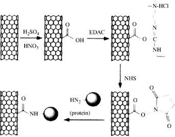
Schematic of the attachment of proteins onto carbon nanotubes through diimide-activated amidation. Reproduced from (14) with permission from Elsevier.
Protein functionalized CNTs for medical applications
CNTs have been functionalized using various proteins, including bovine serum albumin (BSA), DNA, and other proteins. The functionalization or immobilization of proteins onto carbon nanotubes is widely considered to be through covalent linkages. Functionalizing CNTs with proteins will provide various reactive groups, including hydroxyl, amines, thiols, and carboxylic acids (17). Some of the proteins that have been immbobilized onto CNTs through covalent linkages include chrymotrypsin, ferritin, fibrinogen, hemoglobin, and streptavidin. Several researchers have reported the covalent immobilization of proteins onto CNTs using 1-ethyl-3-(3-dimethylaminopropyl) carbodiimide as the crosslinking agent (16). Based on confocal images, Hazani et al have reported that DNA-functionalized SWCNTs had minimal nonspecific interactions with noncomplementary sequences, demonstrating that the majority of the binding was through covalent crosslinkings (18). Although Hazani et al claim to have demonstrated that covalent crosslinking is predominant, Gao et al proposed that immobilization could also be due to absorption rather than covalent crosslinking, because absorption has strong affinity and spontaneity over covalent crosslinking (16).
In one of the earliest studies on functionalizing MWCNTs, Chen et al used 1-pyrenebutanoic acid succinimidyl ester to irreversibly attach onto to the surface of the CNTs through noncovalent interactions (19). This leads to the formation of succinimidyl ester groups that have high reactivity to primary and secondary amines and can be used to attach various types of biomolecules onto the functionalized nanotubes. A schematic of the functionalization of the nanotubes and subsequent attachment of biological molecules through amidation is shown in Figure 2 (19). The potential of immobilizing proteins such as ferritin and streptavidin and smaller molecules such as biotin-PEO-amine was studied. It was reported that a controlled, nanotube-specific method was developed to immobilize proteins and biomolecules as well as inorganic nanoparticles.
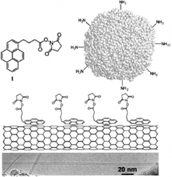
Schematic of the functionalization of carbon nanotubes through amidation. Bottom image is of the single-walled nanotube grown on a gold template. Reproduced with permission from (19): Chen RJ, Zhang Y, Wang D, Dai H. Noncovalent sidewall functionalization of single-walled carbon nanotubes for protein immobilization. J Am Chem Soc. 2001;123(16):3838-3839. Copyright 2001 American Chemical Society.
Protein nanotube conjugates were produced by covalent linkages between SWCNTs and MWCNTs and bovine serum albumin (20). CNTs were first purified by refluxing in nitric acid solution and later attached to BSA via diimide-activated amidation reaction. Although BSA is colorless in solution, a dark color solution was obtained after reaction between BSA and the SWCNATs and MWCNTs, suggesting functionalization. The atomic force microscopy (AFM) image in Figure 3 shows that the proteins are immobilized along the length of the nanotubes. The MWCNTs had a considerably higher attachment of proteins than the SWCNTs due to the presence of a higher number of defects (20). However, functionalizing of SWCNTs was found to be more effective than in MWCNTs because of the resistance offered by the latter due to multiple layers and the larger outer diameter (21).
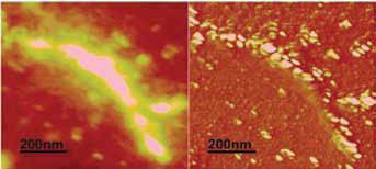
Atomic force microscopy (AFM) images shows that the proteins are immobilized along the length of the carbon nanotubes. Reproduced with permission from (20): Huang W, Taylor S, Fu K, et al. Attaching proteins to carbon nanotubes via diimide-activated amidation. Nano Lett. 2002;2(4):311-314. Copyright 2002 American Chemical Society.
Amino functionalization of SWCNTs was done using 2 approaches based on chemical modifications of the carboxylic groups via amide formation (1). The first approach was to directly couple ethylene diamine with the carboxylic groups to introduce amino groups. The second approach was to reduce the carboxyl group to hydroxymethyl, later transforming the aminomethyl group. A schematic of the 2 approaches is shown in Figure 4. Fourier Transform Infrared Spectroscopy (FTIR) and X-ray photon spectroscopy confirmed conversion of the carboxylic acid to the functionalized SWCNTs. The functionalized nanotubes now had covalently bonded amine groups on the surface that could be used to bind polymer or biological entities for various applications (1). In another study, amino functionalization of SWCNTs was done by covalently attaching N-alkylidene amino groups to the side wall of the CNTs through C-N bonds. The functionalized CNTs were considered useful to further form peptide linkages for the preparation of nylon-type crosslinked SWNTs and covalent sidewall bindings suitable to attach polymer matrices or DNA (3).
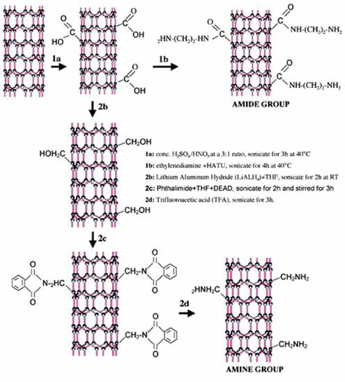
Approaches used to functionalize the carbon nanotubes through a 2-step process. Reproduced with permission from (1): Ramanathan T, Fisher FT, Ruoff RS, Brinson LC. Amino-functionalized carbon nanotubes for binding to polymers and biological systems. Chem Mater. 2005;17(6):1290-1295. Copyright 2005 American Chemical Society.
CNTs were made water-soluble by functionalizing with amino acids (22). Amino acid 116 and paraformaldehyde were added into a dimethylformamide solution containing the NTs and the mixture was heated at 130°C for 96 h. About 10% of the nanotubes were functionalized after the treatment. Later, the fCNTs were treated to remove the n-tert-butoxycarbonyl protecting group. The SWCNTS and MWCNTs obtained with this approach were highly stable in water for up to 30 days. TEM images showed individual SWCNTs with diameters between 20 nm to 30 nm and bundles of SWCNTs with diameters in the range of 10 nm to 50 nm. The amount of amine functional groups was between 0.25 mmol and 0.5 mmol per gram of material (22).
CNTs have been functionalized with various types of proteins and molecules to enable them to penetrate into cells and deliver payloads. Some of the structures that have been proven to be capable of penetrating the cells and that display biological functions are depicted in Figure 5 below (23). CNTs are envisaged to enter into cells (internalize) primarily along 2 pathways. Functionalized nanotubes could enter cells through passive diffusion and cross the lipid bilayer or by endocytotsis after they attach onto the surface of the CNTs (23).
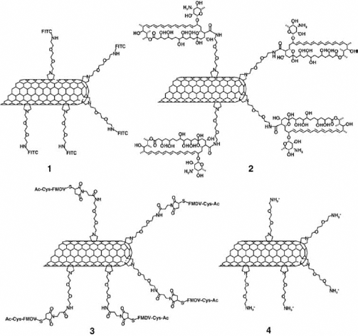
Biological structures that have been proven to be capable of penetrating into cells and retaining biological functions. Reproduced from (23) with permission from Elsevier.
Amino-functionalized CNTs have been used as delivery vehicles for DNA for targeted gene delivery (24). The interaction between plasmid DNA and single-walled and multiwalled amino functionalized CNTs and lysine functionalized SWCNTs (20-70 nm) were studied. Figure 6 shows a SEM image of the MWCNTs attached on carboxymethylated mica surface. Interactions of single and multiwall CNTs functionalized using ammonia and lysine with plasmid DNA were studied (24). Functionalized nanotubes were dissolved in aqueous solutions and later mixed with various ratios of plasmid DNA at room temperature to form complexes. It was concluded that the multiwalled CNTs functionalized with ammonia could bind significantly higher amounts of DNA (due to greater cationic surface density and increased surface area) compared to the SWCNTs functionalized with lysine and ammonia whereas the SWCNTs functionalized with only ammonia did not show any binding of DNA. MWCNTs were also found to have more compact DNA than SWCNTs (24).
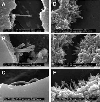
Scanning electron microscopy (SEM) picture showing the attachment of DNA onto the single-walled carbon nanotubes and multiwalled carbon nanotubes at 3 different magnifications. Reproduced with permission from (24): Singh R, Pantarotto D, McCarthy D, et al. Binding and condensation of plasmid DNA onto functionalized carbon nanotubes: toward the construction of nanotube-based gene delivery vectors. J Am Chem Soc. 2005;127(12):4388-4396. Copyright 2005 American Chemical Society.
CNTs were covalently linked with DNA in a multistep approach. Chemical modification (oxidation) was first done to obtain amine terminated CNTs, which were later covalently bound with DNA (25). X-ray photoelectron spectroscopy was used to confirm formation of the amine terminated CNTs. Further, AFM studies conclusively showed that the chemical functionalization occurred at the ends and sidewalls of the CNTs, as seen from Figure 7.
Atomic force microscopy images show that chemical functionalization and attachment of DNA occurred at the sidewalls (
A noncovalent binding of unmodified siRNAs on to SWCNTs for efficient transport into cells and the liberation of siRNA for efficient gene silencing was developed by Krajcik et al. SWCNTs were first oxidized and later functionalized with hexamethylenediamine (26). To prepare siRNA-SWNT complexes, aqueous solutions of functionalized SWCNTs were combined with siRNA, ensuring that no agglomeration occurred. It was concluded that SWCNTs functionalized with HMDA and poly(diallyldimethylammonium)chloride (PDDA) were found to be suitable for forming complexes with siRNA where the siRNA-SWNT complexes were able to translocate into primary cells. The siRNA was released and induced efficient silencing of target genes. SWCNTs were functionalized with hexamethylenediamine (HMDA) and poly(diallyldimethylammonium)chloride (PDDA) that was able to bind the negatively charged siRNA through electrostatic interactions. No cytotoxic effects were observed even at a nanotube concentration of 10 mg/l and the functionalized CNTs were able to cross the cell membrane (26).
The ability of SWCNTs and MWCNTs to load plasmid DNA and enter cells, together with the extent of influence on gene expression, was investigated by Pantarotto et al. In this study, CNTs were functionalized by adding a pyrrolidine ring with an amino-terminal oligoethylene glycol attached to a nitrogen atom through covalent bonding. Concentration of the functional groups was estimated to be about 0.55 mmol/g and 0.90 mmol/g for the SWCNTs and MWCNTs, respectively. After loading with plasmid DNA, the CNTs were incubated with HeLa cells at a concentration of 2.5 mg/ml. Figure 8 shows a TEM image of the HeLa cells after incubating with the CNTs. Nanotubes inside the cells were found to have a diameter of about 20 nm and length of about 200 nm, similar to the original dimensions of the nanotubes. This suggests that the nanotubes entered the cells as nanoneedles without causing cell death (27). No cytotoxicity was observed even when the concentration of the nanotubes was as high as 1.2 mg/ml.
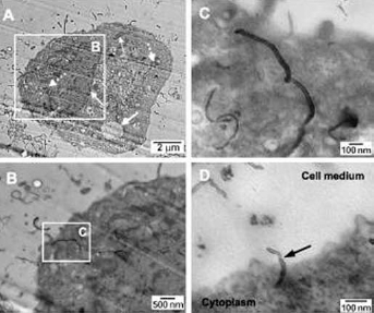
Images of HELa cells containing the carbon nanotubes. (
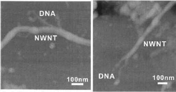
Higher gene expressions (5-10 times) have been observed in cells containing CNTs compared to those without nanotubes. Recent studies have shown that pyrimidine functionalized CNTs incubated with single-stranded DNA formed macroscopic aggregates that were radially bound to form a 3D lattice structure, as seen in Figure 9 (28, 29). DNA was used to effectively disperse and separate bundled SWCNTs in water (28). Based on molecular modeling studies, it was suggested that single-stranded DNA could bind to CNTs through π-stacking, resulting in helical wrapping on the surface. Further, the DNA dispersed CNTs were separated according to their electronic properties (29) and multi-step approach was used to covalently link MWCNTs to DNA. Oxidized CNTs having open ended nanotubes with terminal carboxylic groups were loaded with DNA and it was found that DNA could be attached at both the sidewalls and ends as described earlier. Since it is difficult to visualize DNA using microscopes, Guo et al introduced several heavy atoms into the double strand of DNA and attached the modified DNA onto CNTs (30). The heavy atoms increased the electron scattering and the image resolution enabling visualizing of DNA attached on to CNTs as shown in Figure 10.
3D lattice structure formed from the macroscopic aggregates of pyrimidine functionalized carbon nanotubes containing single-stranded DNA. Reproduced with permission from (28): Arnett CM, Marsh CP, Welch CR, et al. Enzyme-mediated assimilation of DNA-functionalized single-walled carbon nanotubes. Langmuir. 2010;26(2):613-617. Copyright 2010 American Chemical Society.

Transmission electron microscopy (TEM) image of a carbon nanotube showing the attachment
Covalently linked peptide nanotubes were functionalized to obtain wires containing free amino groups (31) with about 0.3 mmol/g to 0.5 mmol/g of functional groups loaded per gram of the NTs. Further structural analysis showed that the nanotube peptide assumed the secondary configuration necessary to be recognized by specific antibodies and the NTs were considered to be suitable to modulate ligand-receptor interactions. SWCNTs dispersed using a 29-residue peptide were used to demonstrate that the peptide organizes the SWCNTs into fibrous arrays (32). Using specific sonication (1 or 4 min, 10 W) and centrifugation (successive centrifugation at 20 000 g for 15 min, 50 000 g for 30 min and 100 000 g for 1 h), it was demonstrated that individual peptide-wrapped SWCNTs could assemble into longer structures (1.2 ± 1.1 µm in length and average diameter of 2.4 ± 1.3 nm) through peptide-peptide interactions. Figure 11 shows an AFM image of the assembled CNTs (32).
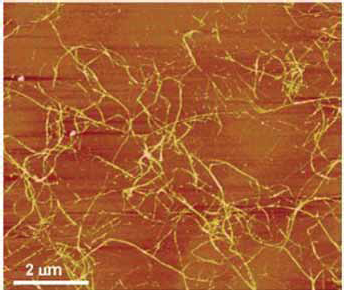
Atomic force microscopy (AFM) image of the single-walled carbon nanotubes assembled into long structures. Reproduced with permission from (32): Zorbas V, Ortiz-Acevedo A, Dalton AB, et al. Preparation and characterization of individual peptide-wrapped single-walled carbon nanotubes. J Am Chem Soc. 2004;126(23):7222-7227. Copyright 2004 American Chemical Society.
SWCNTs and MWCNTs were functionalized using bovine serum albumin using carbodiimide-activated amidation reaction (20). In a similar approach, CNTs functionalized using oligomeric polyethylene glycol compounds that contained ester linkages were later used as starting material in ester-to-amide transformation reactions with BSA proteins (33). An investigation was done to understand the binding of BSA to MWCNTs functionalized with 4 different chemicals that differed in their length, diameter, and structure (34). Using steady-state and time-resolved fluorescent experiments, it was demonstrated that the proteins were bound onto the CNTs by forming an complex. In addition, it was found that nanotubes with larger diameters had stronger protein binding compared to smaller diameter nanotubes. Negative charges or steric properties enhanced binding for some proteins and not others suggesting that electrostatic and stereochemical interactions of the nanotubes and the proteins played a part in determining protein binding (34).
Bovine serum albumin (BSA) and other proteins were used to solubilize SWCNTs using a simple approach (17). Aqueous dispersions of SWNTs were prepared by dispersing in dimethyl formamide. Later, the nanotubes were dispersed in protein solutions and ultrasonicated. Figure 12 shows digital images of the SWNTs dispersed in water with and without BSA. As seen from the figure, the addition of BSA substantially improved the solubility; the inclusion of Mucor javanicus lipase further increased the solubility. These CNT-protein solutions were found to be stable without any aggregation even after 30 days. Atomic force microscopy (AFM) images showed that the CNTs were individually dispersed with an average diameter of 1.2 ± 0.4 nm. It was suggested that intense ultrasonication frayed the ends of the SWNTs and exposed them in solution. Proteins in solution were able to adsorb onto these frayed ends through hydrophobic interaction, π-π interactions, or through amine functionalities, making the hydrophilic/charged group interact with water and the nanotubes soluble (17). Functionalization of double-walled CNTs (DWCNTs) with BSA increased dispersion and AFM studies showed that BSA was able to attach as single molecules onto the surface. Based on the differences in height of the DWCNTs before and after functionalization, it was observed that biotin-BSA-functionalized CNTs could specifically bind to streptavidin, demonstrating noncovalent binding. Uniform and dot-like distribution of streptavidin was seen on the biotin-BSA-functionalized nanotubes. Such specific binding and recognition of biomolecules will be useful for detection of various proteins and polymers (35). Amino-functionalized CNTs were treated with BSA and DNA and the attachment of these biomolecules was observed using transmission electron microscopy (TEM) and Fourier transform infrared spectroscopy (FTIR). Amino functionalization decreased the length of the MWCNTs from 30-80 µm to 10-20 µm and substantially increased the dispersion, but made the surface of the nanotubes rougher. Uniform attachment of BSA on to the surface of the fMWCNTS can be seen from the TEM images in Figures 13 A and B. Further functionalization of the CNTs with DNA showed similar attachment onto the surface (36).
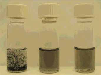
Differences in the solubility of the carbon nanotubes without
Transmission electron microscopy (TEM) images of BSA (globular and elongated shape) attached onto amino-functionalized multiwalled carbon nanotubes at 2 different magnifications. Reproduced from (36) with permission from Elsevier.
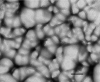
In a recent study, a commercially available mixture of soy protein and milk protein was functionalized onto MWCNTs using a 2-step MWCNT fabrication process (37). In the first step, the protein and metal salt was prepared and dried; then, in the second step, the dried materials were decomposed in a furnace to form the CNTs. Later, the MWCNTs were purified to remove volatile contents and metals and were used for further analysis. Based on TEM and X-ray diffraction (XRD) results, it was found that the nanotubes obtained were about 100 nm in size and structurally polycrystalline.
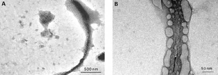
Feritin, a protein from horse shell that contains about 24 units with diameters up to 12 nm to 13 nm was used to study the affinity of protein to SWCNTs in water (38). Up to 25% of the feritin was solubilized and was well dispersed, as shown in Figure 14A (38). The better dispersion was suggested to be due to the nonspecific interactions, including electrostatic interactions, hydrophobic interactions, and hydrogen bonding. Addition of activation agents such as 1-Ethyl-3-(3-dimethylaminopropyl)carbodiimide) (EDAC) promoted conjugation of the ferritin proteins through covalent linkages. The TEM image in Figure 14B shows that the EDAC-containing solution had a considerably higher number of SWCNTs. It was concluded that SWCNTs had a natural affinity to proteins when in an aqueous solution but the protein affinity can be controlled or eliminated by covalent functionalization of the nanotubes with hydrophilic polymers or with oligomeric PEG (38).
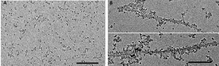
Changes in the dispersibility of feritin in water without (
Natural proteins such as lysozymes have also been functionalized onto CNTs for biosensing and other applications using various approaches. Lysozymes are presumed to be adsorbed onto CNTs through hydrophobic and π-π interactions. Lysozyme and oxidized CNTs were found to mainly have electrostatic interactions. The amount of lysozyme attached onto the CNTs was dependent on the pH and could be controlled by varying the net charge (39, 40). Denaturation of lysozyme was not observed during the interaction of the lysozyme with CNTs. It was suggested that covalent binding may be necessary to retain the lysozyme for rigid applications (39, 40). Four proteins (fibrinogen, c-globulin, hemoglobin, and fibronectin) were used as intermediates for self-assembly of gold nanoparticles onto functionalized MWCNTs (41). It was suggested that the protein-assisted binding of metallic nanoparticles could be used to develop CNT-based catalysts for supercapacitors and various applications. Lipases extracted from Candida rugosa were covalently anchored onto acid-treated MWCNTs through a self-catalytic mechanism that made the CNTs soluble in organic solvents such as tetrahydrofuran (THF), dimethylformamide (DMF), and chloroform (42). Unlike the unmodified CNTs that precipitated in these solvents, lipase-modified CNTs were stable for 2 weeks without any precipitation, which enables the use of the modified CNTs for biosensor and biocatalyst applications.
Understanding the interaction of blood proteins with CNTs would be beneficial to design CNTs for various biomedical applications. In a detailed study on the interaction of the blood proteins Bovine Fibrinogen (BFG), Gamma globulin (Ig), Transferrin (Tf), and bovine serum albumin (BSA), it was found that the binding of proteins onto CNTs was determined by the structure, amino acid composition, and molecular weight, among other factors (43). AFM images of CNTs after incubating in various proteins after 10 min and 5 h is shown in Figure 15. BFG and Ig were attached onto the CNTs in higher quantities due to the large number of hydrophobic regions and presence of tryptophan, tyrosine, and phenylalanine, which contain aromatic rings that can facilitate attachment of CNTs through π-π interactions. These 2 proteins attached onto CNTs with a height of about 15 nm to 30 nm, compared to less than 20 nm for Tf and BSA. However, Tf and BSA were able to assemble and reach thermodynamic equilibrium comparatively quickly and no significant difference was seen in AFM images of CNTs incubated with these proteins after 10 min and 5 h (43). The amount of proteins adsorbed onto CNTs was also found to be highly related to the molecular weight or size of the proteins. For instance, BSA, which had a dimension of 13.2 × 8.5 × 7 nm, compared to 25.3 × 6.5 × 53.5 nm for BFG, had much lower adsorption. Longer proteins would have a higher number of binding sites and consequently the chance for better attachment (43). Considerable changes in the α-helix and β-sheet contents were also observed depending on the type of protein and amount of protein adsorbed.
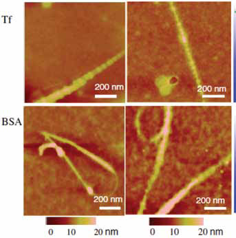
Atomic force microscopy (AFM) images of blood proteins (Tf, BSA) attached onto the carbon nanotubes 10 min and 5 h after incubation. Reproduced from (43): Ge C, Du J, Zhao L, et al. Binding of blood proteins to carbon nanotubes reduces cytotoxicity. Proc Natl Acad Sci USA. 2011;108(41):16968-16973, with permission from the National Academy of Sciences of the USA.
Although most proteins have been successfully attached onto CNTs through various physical and chemical means, there are several pre-requisites for proteins to attach onto CNTs. For example, amphiphilicity was required for proteins to disperse CNTs and only amphiphilic proteins containing hydrophobic residues at the center or end of the sequence were able to provide stable dispersions. Similarly, aromatic amino acids were found to be required for the binding of peptides onto CNTs. Four polypeptides with a particular aminoacid sequence were reported to be responsible for protein binding onto CNTs (44). The strength of the binding between CNTs and peptides was directly dependent on the number of interacting residues on the surface of the peptides (45). It is necessary to thoroughly understand the structure, properties, and behavior of the proteins and their interaction with CNTs to use protein-functionalized CNTs for specific applications.
Protein-functionalized CNTs as biosensors
Functionalized CNTs can be used as biosensors because the nanostructure of CNTs together with their conducting properties enable tiny signals to be detected and transmitted that help locate target molecules at low concentration levels (16). CNTs functionalized with various proteins have been studied for potential biosensing applications. Some of the proteins studied for functionalization of CNTs intended for biosensing applications include acetylcholinesterase, avidin, BSA, chymotrypsin, cytochrome C, ferritin, lipase, lysozyme, and streptavidin as seen from Table I. In 1 study, calf thymus DNA was immobilized on the surface of MWCNTs for potential use as DNA biosensors (46). Cyclic voltammetry and electrochemical impedance analysis showed that peak current decreased and the electron transfer resistance increased after immobilization through diimide-activated amidation. However, absorption of the DNA onto the surface of the CNTs was also not ruled out. Interactions between DNA molecules immobilized on the CNTs with small biomolecules such as ethidium bromide was observed, indicating that the DNA molecules had undergone minimum structural change and could interact with other biomolecules (46).
Some of the proteins used to functionalize carbon nanotubes and their potential applications
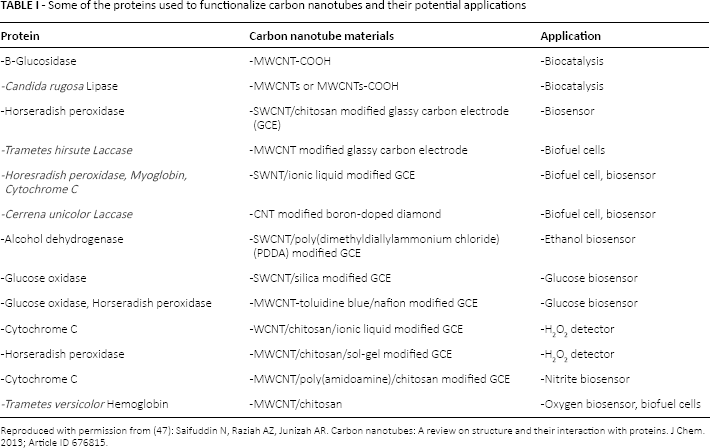
Reproduced with permission from (47): Saifuddin N, Raziah AZ, Junizah AR. Carbon nanotubes: A review on structure and their interaction with proteins. J Chem. 2013; Article ID 676815.
Oxidized SWCNTs were functionalized with knob protein domain from adenovirus serotype 12(Ad 12 knob) or its human cellular receptor, the coxsackic virus and adenovirus receptor (CAR) protein via diimide-activated amidation (11). AFM images showed that the protein complexes were attached along the length of the SWNT. The functionalized CNTs retained their biologically active conformation and current-gate voltage measurements showed that the modified CNTs could act as biosensors, detecting protein activity and environmental adenoviruses (11).
A biofunctional molecule 1-Aminopyrene (1-AP) containing a pyrenyl group and an amino functional group was used to make MWCNTs disperse in aqueous solutions and also to assist in enzyme immobilization for potential use as biofuel cells (48). Laccase was immobilized on MWCNTs functionalized with 1-AP using glutaraldehyde crosslinking. Functionalizing MWCNTs with 1-AP made the MWCNTs easily dispersible in an acetate buffer solution, as seen in Figure 16. Compared to unfunctionalized MWCNTs, the 1-AP functionalized MWCNTs had higher catalytic activity and better stability with a current density of about 21.4 A/Cm2 towards oxygen reduction at pH 7, demonstrating the usefulness of the MWCNTs as biofuel cells.
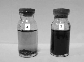
Digital image of pristine carbon nanotubes (
Amino-functionalized CNTs treated with iron phthalocyanine were investigated as a catalyst for the oxygen reduction reaction (ORR) in an air cathode single cell microbial fuel cell (49). Considerably high electrocatalytic activity (power density of 601 mWm2) was obtained using the amino-functionalized CNTs.
Functionalizing of enzymes onto CNTs has been performed using various approaches. Bioelectrodes for sensing glucose and ethanol were fabricated by wrapping single-stranded DNA on to the CNTs and immobilizing enzymes onto the DNA-wrapped CNTs (50). The enzyme-immobilized CNTs had increased activity and stability of glucose oxidase and laccase, while power production was also enhanced (50). Surfactants such as Triton X-100 were found to specifically bind streptavadin and facilitate interfacial electron transfer of the proteins with enhanced faradic responses. In another study, a layer-by-layer approach has been adopted to immobilize various types of enzymes onto CNTs. This approach enables increasing the biocatalytic activity of the CNTs to the desired level by increasing the number of enzyme layers (51). Strong electrostatic interactions between the DNA and protein in addition to van der Waals and π-π interactions provide excellent adhesion of the enzymes to the CNTs. These fCNTs were found to have ultrasensitive detection that was considerably higher that common sensors, leading to high levels of DNA or protein detection (51). Rather than using physical absorption, which could cause leaching and durability issues, covalent binding of enzymes has also been performed. Enzyme loadings as high as 1000 µg/g of CNT have been obtained through covalent crosslinking (51).
Cytotoxicity of protein functionalized CNTs
Potential cytotoxicity of CNTs is 1 of the major limitations for medical applications. However, contradictory reports have been published on the cytotoxicity of CNTs. It has been suggested that cytotoxicity of CNTs is dependent on the concentration of the CNTs in the cells, the cell types, and thetype and level of functionalization (52). The 4 different blood proteins discussed earlier had lower cytotoxicity than uncoated CNTs. Among the 4 proteins, BFG-coated CNTs showed no toxicity, which was attributed to the multi-layer arrangement of the proteins on the surface and the covering of the CNT surface, thereby avoiding exposure of the CNTs to cells. However, CNTs containing high levels of BFG showed lower cell viability compared to those with alower level of loading (53). BSA was shown to disperse and BSA-dispersed CNTs were readily taken up by HeLa and human mesenchymal stem cells (hMSCs) with distinct subcellular localization even in the cytoplasm. As high as 86 ± 33 x106 and 21 ± 33 x106 of SWCNTs were detected per hMSC and Hela cell, respectively, without any adverse effects to the cells (54).
Conclusions
The functionalizing of CNTs with proteins has mutual benefits that enable the unique properties and application of CNTs to be realized for various fields. CNTs have been functionalized with various proteins and used predominantly for medical applications, including biological sensing. Protein-functionalized CNTs have shown potential for having high pay loads and long release rates. The functionalized CNTs also retain their biological function and enter cells relatively much easier than free proteins. In addition to conventionally used proteins such as BSA, collagen, and fibrinogen, plant proteins such as zein and soy proteins should be considered for functionalizing the CNTs. Although a couple of reports are available on the use of plant proteins such as soy proteins for functionalizing CNTS, further research is necessary to understand the potential of using cereal proteins to functionalize CNTs for various applications. Until now, protein-functionalized CNTs have mainly been studied for biosensing, biocatalysis, and medical applications. New application areas in food, energy, and electronics should be explored for CNTs with or without functionalization. Although most studies show that CNTs at low concentrations do not affect cytocompatibility, specific studies do demonstrate cytotoxic effects of CNTs. Further research is necessary to understand the cytotoxic and carcinogenic effects of CNTs before and after functionalization with proteins.
Footnotes
Financial support: Financial support to complete the work was received from the Ministry of Science Technology, Department of Biotechnology, Government of India through the Ramalingaswami Fellowship to NR, and from the Center for Emerging Technologies, Jain University.
Conflict of interest: The authors declare no conflict of interest.