
Abstract
Introduction
The optical properties of dental restorative materials have a dramatic effect on patient esthetics, which may be compromised by the poor blending effect of composites resins at the composite–tooth interface.
Objective
The objective of this study was to evaluate the color-matching ability and the blending effects of 3 different composite resins when restoring natural teeth.
Methods
Three commercially available composites and 60 central incisors were used for this experiment. Each tooth was sectioned horizontally at the level of the cementum–enamel junction, and the crown was then bisected along the long axis of the tooth. One half of each tooth was restored individually with composite resin, after matching with the corresponding tooth shade. The tooth color was evaluated preoperatively and postoperatively using an intraoral spectrophotometer and a scientific spectrophotometer. Color differences were then evaluated by the CIEDE2000 color difference formula.
Results
The results showed the existence of color differences between the intact and the restored sections of the teeth. However, these differences were considered acceptable, since the ΔΕ value ranged below 3.3. The ΔE, Δa and Δb parameters showed no statistically significant differences between the groups (p>0.05).
Conclusions
The 3 composites tested were able to mimic natural teeth and produce acceptable restorations. However, the color of the resin composites needs to be evaluated over the long term, because it is subject to alterations in the oral environment over time.
Introduction
One of the most challenging tasks a dentist has to confront in daily practice is matching the color of composite restorations used in anterior teeth. Manufacturers have produced an array of dental composites that were initially introduced as wear resistant, with excellent optical properties and high polishing capabilities. Matching the color of a composite restoration, as well as the reproduction of an “intact” tooth with the use of resin composites, however, constitutes a very challenging task (1, 2). Moreover, discrepancies between shade guides and the actual shade of the composites have been recorded (3). Thus, the use of digital color matching instruments has become part of dental education, while the Vitapan Classical shade guide has become the most popular shade guide (4).
The fillers used in composite resins significantly influence the close-to-natural visual outcome and the blending effect – which is essential to meet the esthetic demands placed on composite resins – as well as the longevity of the restorations. The size, shape, content and refractive index of the fillers contribute to the color outcome of composites (5). Besides the fillers, the optical properties of composite resins are also determined by the type of monomers in the resin matrix. The amount of Bis-GMA contained in the composite also has an effect on the translucency and the refractive index of composite resins and is an alternative way, used often by manufactures, to adjust the optical properties of composite resins (6).
There are several factors that render the color matching difficult. These difficulties arise from the fact that color matching depends on several different chromatic properties associated with teeth and composite resins. Those properties include hue, value and chroma; translucency, fluorescence and opalescence; light transmission and diffusion; and texture and luster of the surface (7, 8). It should be noted that the color of the teeth is mainly determined by the dentin and not the enamel, with the latter having a minor influence on the color of the teeth (9, 10) but a main influence on the color perception in terms of lightness. However, the simulation of the dental tissues has been made possible due to the natural layering concept. In this concept, the missing dental tissues are replaced in increments with composite resins of the exact shade as the dental tissue. A translucent composite resin is applied over a more opaque composite resin to achieve a depth perception similar to that of the natural teeth (11-12-13).
Clinically relevant color difference parameters include the magnitudes of total color difference (ΔΕ) that is both perceptible and acceptable to human observers. Perceptibility refers to the detection of the color difference between a tooth and an adjacent colored restoration, whereas acceptability refers to whether the color difference would be acceptable for the restoration (14). One of the color measurement methods in dentistry is the CIEDE2000 total color-difference formula, according to which color changes are considered to be visually perceptible when ΔΕ >1, and clinically acceptable when ΔΕ <3.3 (15).
The aims of this study was to evaluate the color-matching ability and the blending effect of 3 different composite resins when restoring the natural teeth. The null hypothesis tested was that there were no color differences (ΔΕ <3.3) between the intact tooth substrate and the tooth restored with the 3 composite resins tested independently.
Methods
The study protocol was reviewed and approved by the ethics committee of Aristotle University of Thessaloniki (protocol No282-13/2/2012). Teeth were extracted for periodontal reasons, and patients’ consent was obtained prior to the study.
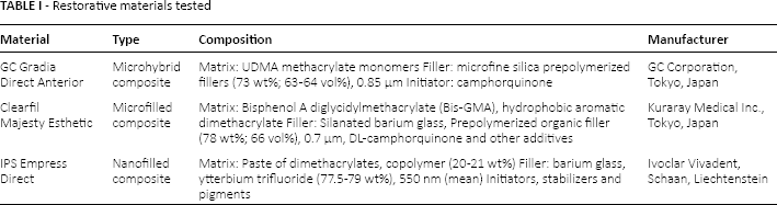
Sixty freshly extracted anterior teeth without any restorations (30 maxillary central incisors, 30 mandibular central incisors) and 3 representative composite resins for anterior restorations (microhybrid, microfilled and nanofilled) were selected. The manufactures, types and compositions of the tested materials are presented in Table I. The teeth were scaled and polished using a rubber cup and pumice. Decayed and discolored teeth were excluded from the study. Prior to the baseline color measurement, the teeth were immersed in distilled water and stored in the deep freezer for 24 hours (-10°C). This procedure was followed to avoid changes in the optical properties of the teeth, because other storage media, such as thymol, have been shown to cause such changes (16, 17).
Restorative materials tested
Each tooth was sectioned horizontally at the level of cementum–enamel junction using a 0.3-mm thick diamond-coated low-speed band saw (Isomet 1000; Buehler, Lake Bluff, IL, USA) under copious water coolant. After sectioning, 2 impressions (double mix impression technique) of each crown were produced using a silicone impression material (Panasil tray soft and Panasil initial contact light; Kettenbach GmbH & Co, KG, Kettenbach GmBH & Co, Eschenburg, Germany USA). The first impression served as a silicone matrix for the specimens to be reproduced with same anatomy as the original teeth. The second impression served as a mold, which allowed repeated measurements of the crown in exactly the same position in the spectrophotometer, which was calibrated at the beginning of the measurements.
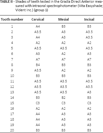
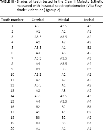
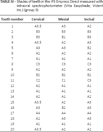
The shade of each tooth was measured in the cervical, middle and incisal third using a intraoral spectrophotometer (Vita Easyshade; Vident Inc., Brea, CA, USA) and evaluated visually with the Vitapan Classical shade guide. The shades of the teeth and materials are summarized in Tables II-IV. The crown was then inserted into the scientific spectrophotometer (Shimadzu UV-2401PC Series, UV-VIS), and 3 consecutive readings were taken for each crown specimen; L*, a* and b* parameters were produced, and a mean value of the readings was calculated (18).
Shades of teeth tested in the Gradia Direct Anterior measured with intraoral spectrophotometer (Vita Easyshade; Vident Inc.) (group 1)
Shades of teeth tested in the Clearfil Majesty Esthetic measured with intraoral spectrophotometer (Vita Easyshade; Vident Inc.) (group 2)
Shades of teeth in the IPS Empress Direct measured with intraoral spectrophotometer (Vita Easyshade; Vident Inc.) (group 3)
Subsequently the crown of each tooth was bisected along the tooth axis, using a 0.3-mm thick diamond-coated low-speed band saw (Isomet 1000; Buehler, Lake Bluff, IL, USA) under copious water coolant. One half of each tooth was then restored with a composite resin of the same shade, as determined earlier with the help of the silicone matrix, thus producing 60 restored tooth specimens. In all of the specimens, each increment of composite resin was placed sequentially from the cervical to the incisal third. This approach has the advantage of masking the interface between the tooth and the restoration and reproducing the optical properties of each of the missing dental tissues (19). The margins of the cavity preparation were not beveled, and the composite was built using the natural layering concept (20); each increment of composite was 1-mm thick and was light cured with Bluephase LED curing light (1,200 mW/cm2; Ivoclar Vivadent AG, Schaan, Liechtenstein) for 30 seconds. The average estimated surface area was 117 mm2 for each cavity (21). The shade of each brand of composite resin was selected such that it exactly matched the shade of the tooth. The focus of the operator was to reconstruct the original anatomy of the tooth with the help of the silicone matrix, thus the filling method was not standardized. The restored specimens were then polished with Super-Snap (Shofu Inc., Kyoto, Japan) starting with coarse discs and ending with extra-fine discs. It should be noted that all of the procedures were performed by the same operator. The specimens were carefully removed from the silicone matrix and placed in distilled water at 37°C for 24 hours with 100% relative humidity, to allow the rehydration of enamel and dentin. The number of specimens to be prepared was defined by statistical calculations with reference to a pilot study.
The specimens were then divided into 3 groups based on the manufacturer of the composite resin chosen for the restoration as presented in Table I:
Group 1: Gradia Direct Anterior (GC Corporation, Tokyo, Japan);
Group 2: Clearfil Majesty Esthetic (Kuraray Medical Inc., Tokyo, Japan);
Group 3: IPS Empress Direct (Ivoclar Vivadent, Schaan, Liechtenstein)
Each tooth specimen was measured again using a scientific spectrophotometer (Shimadzu UV-2401PC Series, UV-VIS), against a black background, while the spectrophotometer was calibrated to a white standard which consisted of a pressed powder tablet of barium sulfate according to the manufacturer's instructions.
The total color difference between the intact tooth and the analogous specimen (CIEDE2000) was calculated using the following equation:

where ΔL was the change of lightness, ΔC = difference in chroma ΔΗ = difference in hue; and Rt, Sl, Sc and Sh are weighting factors. The 3 K terms were additional weighting factors that were set to 1.
Statistical Analysis
The Shapiro-Wilk test indicated that the data thus obtained followed a normal distribution. The equality variances across subgroups were verified by Levene's test. The comparisons of mean scores of specimens’ ΔΕ were evaluated with 1-way analysis of variance (ANOVA) and Tukey's HSD post hoc test, while statistical significance for all the tests was set at a p value <0.05. The statistical analysis was carried out using the SPSS 18.00 for Macintosh software package (SPSS Inc., Chicago, IL, USA).
Results
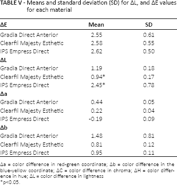
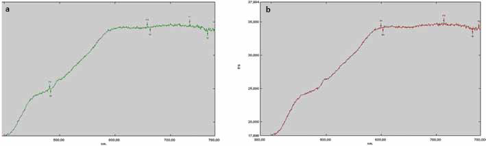
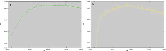
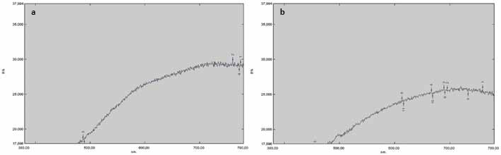
The mean values of ΔΕ, ΔL, Δa and Δb, and standard deviations of each tooth specimen are presented in Table V. Representative figures of spectral reflectance curves are presented in Figures 1-3. These figures represent total color differences between intact teeth and restored teeth. The ΔΕ value between initial and postrestoration values showed that there were total color differences in all groups ranging from: (i) 0.89-3.74 for group 1 (n = 20), (ii) 1.60-3.80 for group 2 (n = 20) and (iii) 1.58-3.30 for group 3 (n = 20). However, the mean value of ΔΕ for all groups was less than 3.3, which indicated that all of the restorations were acceptable to the human eye (11, 15). The ΔΕ, Δa and Δb values among groups were not statistically significant (p>0.05). However, the ΔL value between the Clearfil Majesty Esthetic and IPS Empress Direct groups exhibited statistical significance (p<0.05). Subsequently, the Ivoclar group exhibited the highest mean color-differences value (ΔΕ) compared with the other 2 groups.
Means and standard deviation (SD) for ΔL, and ΔE values for each material
Δa = color difference in red-green coordinate; Δb = color difference in the blue-yellow coordinate; ΔC = color difference in chroma; ΔH = color difference in hue; ΔL = color difference in lightness
p<0.05.
Spectral reflectance curve of intact tooth
Spectral reflectance curve of intact tooth
Spectral reflectance curve of intact tooth
Discussion
The null hypothesis of the present study was accepted since no significant statistical differences in the total color-differences parameter ΔΕ between the groups (p>0.05) were observed. The scientific spectrophotometer calculated the parameters L, a, and b for each tooth specimen before and after the restoration, from which the values of ΔΕ, ΔL, Δa and Δb resulted. The total color difference (ΔΕ) was evaluated for acceptability and perceptibility. The 50%: 50% threshold was set to 1 (ΔΕ <1); such a limit indicates that 50% of observers are able to detect the differences, whereas 50% are not. A color difference above 3.3 was set as the threshold of acceptability for composite resin restorations. However, a color difference that is greater than a ΔE value of 3.3 may still be clinically acceptable, depending on the degree of polychromaticity and translucency of the teeth adjacent to the restoration (22). In our study the ΔΕ value was above the threshold of perceptibility (ΔΕ >1). Therefore, color differences were perceptible, meaning that 50% of observers could detect the difference and 50% could not (23). On the other hand, the ΔΕ value was below 3.3, which constitutes the threshold of acceptability (11, 15).
Østervemb et al (24) evaluated the color-matching ability of composite resins compared with the intact tooth structure and found that the composite restoration was esthetically acceptable via the color differences formulas (CIELab, ΔE2000). They used a specially designed apparatus to cut off a rectangular tooth piece extending from the middle to the incisal third of the tooth crown. Composite systems were used to make the same restoration and the extended visual rating scale for appearance match (EVRSAM) criteria were used to evaluate the esthetic match. The rating scale of the EVRSAM ranges from 0 to 10, where 0 is given when the restoration can be delineated with difficulty and 10 when the restoration has completely unacceptable esthetics. In our study, the crown of the tooth was bisected from the incisal to the cervical third of the tooth, and color differences parameters ΔΕ, ΔL, Δa and Δb were evaluated using a scientific spectrophotometer. Li et al (25) compared natural enamel with 4 translucent composites and found statistically significant differences between intact enamel slabs and translucent composite specimens. Friebel et al (26) also found that composite specimens made up completely of enamel shade showed ΔΕ >4 which is not clinically acceptable. On the other hand, the same authors found that specimens made up of enamel shades in combination with dentin shades in layers of 1 mm each were barely distinguishable (26). However, the authors specify that their study cannot be equated to the color perception of natural teeth and can only be used as an approximate value. This is because the layers of natural teeth have a morphology that is irregular in thickness, with an irregular surface structure (26). In our study, the optical characteristics of enamel shades in combination with dentin shades – of 3 different commercially available composite resins – used in anterior restorations, in the exact morphology of the natural tooth, were compared, thus imitating the clinical process. The restored teeth were fabricated to the exact dimensions of the intact teeth to standardize color measurements and identify total color difference in the same tooth, before and after the restoration. Regarding the parameters ΔL, Δa, and Δb, the results of this study were in accordance with the results of other studies, where the ΔL* parameter did have a statistically significant difference, suggesting that the restored teeth did not have the same lightness value as intact tooth. However, the difference was not noticeable to the human eye (26). The above is justified, because it is harder to perceive a color difference due to lightness between 2 specimens than a color difference due to hue (27). The high values of ΔΕ when pure enamel was compared with translucent composite resins are justified by the fact that the size, orientation and concentration of filler in the resin composites are different from that of enamel prisms, leading to optical differences between them (25). The review of the overall differences in color coordinates between the intact teeth and the restored teeth indicates that the difference in the lightness coordinate ΔL* had a positive value for all groups, which means that there was a shift to a lighter shade. For the groups of Gradia Direct Anterior and Clearfil Majesty Esthetic, the difference in the red-green coordinate (Δa*) had a positive value (+a*), which indicates a shift to red, while for the IPS Empress Direct there was a negative value (-a*), which indicates a shift to green. For all of the groups, the difference in the blue-yellow coordinate (Δb*) had a positive value (+b*), which indicates a shift to yellow.
The differences in the color parameters between the composite resins can be justified based on the different matrix composition. Gradia Direct Anterior is a UDMA-based composite resin, while Clearfill Majesty Esthetic and IPS Empress Direct are Bis-GMA and Bis-GMA with UDMA based, respectively. Bis-GMA has a direct effect on translucency and optical properties, even when the filler content is constant. The color differences between the composite resins with increasing Bis-GMA content were also clinically perceivable (5). The amount of fillers in composite resins also plays an important role in determining the color of composites. The total color difference increases with an increasing amount of fillers, regardless of the shape (27). In our study, the IPS Empress Direct group had a larger amount of fillers compared with the other 2 groups. As a result, the mean ΔΕ value of the IPS Empress Direct group was significantly higher.
This study attempted to determine the total color differences of restored teeth with resin composites. Previous studies have used specimens of several dimensions without considering the fact that the natural teeth have different morphology, irregularities in thickness of enamel and dentin as well as in the surface structure of natural teeth. In our attempt to overcome this problem, we used a silicone matrix to create the restored tooth specimens. We reproduced the dimensions of the original unrestored teeth, which were previously evaluated in terms of color, so that each tooth served as its own control.
With the natural layering concept and the ability of composite resins to blend with the natural tooth structure, clinical practitioners can achieve an acceptable color match. Yet, scientific instruments such as a scientific spectrophotometer can identify color differences undetected by the human eye. A black background was used because it mimics a Class IV restoration, which is the most challenging task. Likewise, the use of a white background mimics a Class V restoration (28, 29).
An important question is whether and to what extent acceptable or even optimum color matching in a laboratory test is a good indicator of similar behavior in clinical practice. Of course, lab conditions differ from real clinic practice, and perhaps it is unrealistic to directly extrapolate these results to in vivo conditions. On the other hand, by knowing the materials’ properties and behavior under certain conditions, the clinician is prepared to determine more accurately the color of the tooth and composite and give the most esthetic results.
Further in vitro and in vivo studies are needed to evaluate the optical properties of the tooth structure as a whole, rather than those of enamel and dentin separately to simulate true clinical conditions. In the final analysis, the evidence indicates the following:
The simulation of clinical conditions and the use of a scientific spectrophotometer allow detection of very small color differences between composite resin and natural teeth.
The color differences (ΔΕ) detected in the materials used were not statistically significant (p>0.05) and were also indistinguishable by simple visual observation.
The specific color parameters (Δa, Δb) showed no statistically significant differences in any of the 3 composite resin materials tested. The parameter ΔL, which indicates the change of lightness in materials, had a statistically significant difference (p<0.05) between groups (Clearfil Majesty Esthetic and IPS Empress Direct).
The technique of natural layering, according to which different shades of resin composites are placed in the cervical, middle and incisal third of a natural tooth appears to be an effective technique for achieving better esthetic results.
Footnotes
Financial support: The authors have not received any financial support.
Conflict of interest: The authors have no conflict of interest.