
Abstract
Aim
Staphylococcus epidermidis is the most common cause of orthopedic infections. Adhesion and biofilm formation on orthopedic implant surfaces play an important role in the physiopathology of these infections. The aim of our study was to evaluate the adhesion of S. epidermidis on the surface of metals usually used in orthopedics.
Methods
Previously sterilized circular metal plates of titanium (Ti), porous titanium (p-Ti), cobalt chromium (CoCr) and stainless steel (SS) were hung completely submerged in a liquid medium with a known concentration of S. epidermidis (RP62A). They were incubated for 1 h or 24 h at 36°C. After incubation, each plate was washed with PBS and sonicated during 5 minutes in 10 mL of saline. Different dilutions were performed and 100 μL from each sample was cultured on agar plates.
Results
26 metal plates were incubated for 1 h and other 55 metal plates for 24 h. The lowest bacterial count (cfu/mm2) at 1 h was observed in CoCr plates while in p-Ti it was 6 times higher. At 24 h the highest bacterial count was observed in SS plates while the lowest in Ti. However, these differences were not statistically significant.
Conclusions
After 1 h and 24 h of exposure, the lowest adherence was observed in CoCr and Ti plates, respectively. However, bacterial attachment occurred with all materials. It is necessary to further investigate new materials able to avoid bacterial attachment.
Introduction
Biofilm formation on orthopedic implant surfaces plays an important role in infections associated with these devices (1). Biofilms are resistant to the immune system (2) and to antibiotics (3-6) and implant removal is necessary in the majority of cases to achieve a high success rate. Coagulase-negative staphylococci and especially S. epidermidis are currently the most common microorganisms isolated in acute and chronic prosthetic joint infections (7, 8) and they are the principal cause of infection persistence and late loosening after acute prosthetic joint infections (9).
Adhesion of planktonic bacteria to the implant surface is the first step in biofilm formation and it is influenced by a great variety of components (10), including implant characteristics (11-14) (e.g., surface charge, microarchitecture, composition, osseointegrative potential), type of microorganism (e.g., presence of flagella, slime production) and environmental parameters (e.g., temperature, flow conditions, chemical characteristics of the medium, presence of antibiotics) (15). The aim of our study was to evaluate the adhesion of S. epidermidis on different metals (titanium, porous titanium, cobalt chromium, and stainless steel) usually used in orthopedics to know if there is any material more resistant to bacterial adhesion.
Material and Methods
Metal plates processing
Metallic discs of 2 mm thickness and 15 mm to 20 mm diameter were obtained from Ames (Barcelona, Spain). The metals were titanium (Ti: Ti-6Al-4V), cobalt chromium (CoCr), stainless steel (SS), and porous titanium (p-Ti) (Fig. 1). The first three metals were obtained from cutting cylindrical solid bars approximately 2 m long.
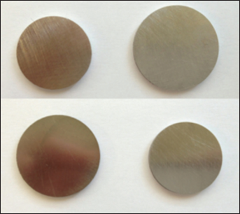
Photo of the four studied metal plates. From left to right and from top to bottom: cobalt chromium, stainless steel, porous titanium and titanium.
The p-Ti was produced by uniaxial compaction of a Ti-6Al-4V powder of grade 5, with an average size of 40 microns. The cylindrical samples were compacted at 600 MPa and sintered at 1300°C for 120 min in a high vacuum furnace with low heating and cooling rates, avoiding oxygen contamination. For the purposes of comparison, all the materials made by the powder metallurgy route were cut and eventually polished, like those coming from a wrought bar. Low porosity level was expected (less than 4 vol.%) with no connection at all. Moreover, after the mechanical treatment of polishing all the remaining porosity was closed (Fig. 2). Hence, identical physical conditions at the surface were promoted in all the samples for the different materials.
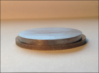
Detail of titanium and porous titanium plates: all the surface of porous titanium was polished, except the edges, where porosity of the material is evident.
Microbiological methods
Staphylococcus epidermidis RP62A (ATCC 35984), was used to perform the study. A colony of RP62A was grown for 24 h at 37°C in Trypticase Soy Broth (TSB, Biomerieux, Lyon, France), then the overnight culture of S. epidermidis was diluted 1:200 in fresh TSB containing 0.25% glucose. Metal discs previously sterilized were completely submerged (Fig. 3) in 100 mL of fresh media with S. epidermidis and then incubated for 1 h or 24 h. After the incubation period, the discs were washed with sterile PBS in order to eliminate all traces of culture media; non-adherent bacteria and discs were then sonicated (Xuba 1, Grant Instruments, Cambridge, UK) for 5 min at 44 kHz in 10 mL of saline serum.
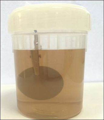
Bottle with a metal plate completely submerged in fresh media with S. epidermidis.
Ten-fold dilutions were performed to quantify the adherent bacteria. Counts were done in blood agar. Results were related to the cfu/mm2 of the metal disc. The experiment was conducted under sterile conditions. A total of 81 plates were processed: 20 SS, 20 Ti, 19 p-Ti, and 22 CoCr.
Statistical analysis
The total cfu divided by the surface of each metal plate (cfu/mm2) was calculated and the variable was log-transformed. We conducted a Kolmogorov-Smirnov test that corroborated the normality assumption of the variable. Finally, we compared the different values of each type of metal at the two different times of incubation (1 h and 24 h) using the ANOVA test. The department of statistics of our center did all the analyses using SPSS v. 16 (SPSS Inc., Chicago, IL, USA) for Windows. Level of statistical significance was established at p≤0.05 (two-tailed).
Results
Twenty-six metal plates were incubated for 1 h and fifty-five metal plates for 24 h. The lowest median bacterial count (cfu/mm2) at 1 h was observed in CoCr plates (1.2787×10² cfu/mm2) and the highest median count was observed in p-Ti (6.6522×10² cfu/mm2), however, the differences were not statistically significant (F=24.34; p=0.131) (Tab. I). After 24 h of exposure, the amount of bacteria attached to the metal surfaces was higher for all metals, the differences between them was lower (F=0.046; p=0.83) and the range of values of bacterial counting increased (Tab. II).
QUANTIFICATION OF S. EPIDERMIDIS AFTER 1 H OF NCUBATION
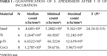
ANOVA test.
QUANTIFICATION OF S. EPIDERMIDIS AFTER 24 H OF INCUBATION
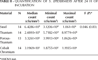
ANOVA test.
Discussion
Prosthetic joint infection is one of the most important problems associated with orthopedic implants. These infections are associated with significant morbidity and with a high economic cost (16-18). Bacterial adhesion and biofilm formation play an important role in physiopathology of these infections, impacting the response in patients treated with debridement and retention of the implant (19-21).
Bacterial attachment to a specific surface depends on bacterial characteristics (specific receptors, external surface net charge) and the characteristics of the colonized surface (chemical structure, electrical charge, porosity) (22-24). In this article, the attachment rate of S. epidermidis to different metals was evaluated. Our results did not demonstrate significant differences in the early or late bacterial attachment to the different metals, only a trend towards lower attachment rate on CrCo after 1 h exposure. These results are in agreement with previous in vitro studies (25) but they are in contrast with animal models in which titanium reduced the infection rate in comparison with stainless steel (26, 27) or a higher inoculum was required to colonize Ti than CrCo (28). A possible explanation for this discordance between in vivo and in vitro studies could be due to the different rate of plasma proteins adhesion to different metals. Plasma proteins mediate bacterial adhesion to artificial surfaces (29, 30).
In conclusion, after 1 h and 24 h of exposure, the lowest adherence was observed in CoCr and Ti plates, respectively. However, differences were not statistically significant and there was bacterial attachment in all experiments, therefore, it is necessary to further investigate new materials that are able to prevent bacterial attachment. More studies are necessary to confirm and corroborate our findings.
Footnotes
Acknowledgements
We would like to thank Dr. José Antonio Calero from Ames S.A., Barcelona, Spain; the staff of the Sterilization Department, the members of the Electromedical Department, Eugenia Guerrero from the Microbiology Laboratory, and Raquel Iniesta from the Department of Statistics at Parc Sanitari Sant Joan de Déu for their wonderful collaboration.