
Abstract
Purpose
We aimed to investigate whether the RANKL/RANK/OPG system is associated with the incidence of periprosthetic osteolysis with septic loosening, and to investigate the differences of RANKL/RANK/OPG system expression in synovial fluid surrounding the normal and septic loosening hip prosthesis in canine models.
Methods
Twelve healthy adult mongrel canines were divided into two groups: experimental and control. Femoral head and stem replacements were conducted on the right side in both groups. The experimental group received the bacteria fluid intra-articular injection and the other group received the same amount of saline in the same day. The synovial fluid samples were gathered at the 1st, 2nd, 4th, 8th, 12th, 16th and 19th week after the bacteria fluid intra-articular injection for enzyme-linked immunosorbent assay (ELISA), the expression of the RANKL/RANK/OPG system.
Results
Surgery on all animals was successful. Two dogs were excluded from the analysis of the result because of a surgery infection or death. The ELISA of the synovial fluid revealed that the ratio of RANKL/OPG showed a significant upward trend (p≤0.05) with time in the test group but the ratio of RANKL/OPG in the control group changed slowly over time (p>0.05). The ratio of RANKL/OPG value between the test and control group showed a significant upward trend, but had no statistical difference (p>0.05) over time.
Conclusions
It could be concluded that the RANKL/RANK/OPG system is correlated with the incidence of periprosthetic osteolysis with septic loosening. Consequently, imbalance RANKL/RANK/OPG system was related to periprosthetic osteolysis with septic loosening.
Keywords
Introduction
Total hip arthroplasty (THA) is one of the most clinically successful and cost-effective healthcare interventions for various types of end-stage hip disorders (1). As the population becomes older, the number of joint prostheses inserted increases steadily (2). A total of 332,000 THA and 719,000 total knee arthroplasty (TKA) were performed in 2010 in the USA. The total numbers are predicted to reach 572,000 and 3.48 million by 2030 for THA and TKA, respectively. In Europe, a larger number of patients underwent primary hip arthroplasty than knee arthroplasty (3–5). While the majority of joint arthroplasties provided pain-free function, a minority suffered device failure and required additional surgeries at some point (6–8). In these cases, one of the main reasons is periprosthetic infection. Using the nation-wide inpatient sample, annual incidence rate of periprosthetic joint infection in the USA increased from 1.99% to 2.18% for THA and from 2.05% to 2.18% for TKA from 2001 to 2009 (9). Once periprosthetic infection occurs, it is very difficult to cure, and will bring tremendous pressure to the patient both physically and psychologically, and it also carries a heavy financial burden for society. The annual cost of infected revisions to US hospitals increased from US$320 million to US$566 million during 2001–2009 and was projected to exceed US$1.62 billion by 2020 (9). During the process of periprosthetic infection treatment, bone deficiency surrounding the prosthesis is a big issue in many cases, which brings difficulties to the revision.
Imbalanced expression of receptor activator of nuclear factor-κB ligand/receptor activator of nuclear factor-κB/osteoprotegerin (RANKL/RANK/OPG) system is closely related to periprosthetic osteolysis with aseptic loosening after THA, which has been confirmed by extensive literature (6, 10–12). However, the pathogenesis of periprosthetic osteolysis with septic loosening is rarely reported. Furthermore, bone deficiency was more common in septic loosening than aseptic, which related to the success of surgery. The result was found when revision of THA was operated from the present study, so we suspect that the RANKL/RANK/OPG system may also play an important role in the pathogenesis of periprosthetic osteolysis with septic loosening (13).
According to An and Friedman (14), dogs have the “closest in vivo condition to the human except for non-human primates.” Factors that make the dog preferable over other higher-level vertebrates (i.e., goats, sheep, and pigs) are the availability of basic data about the species, ease of handling, and modest housing requirements. In studies by Skurla and James, the incidence of aseptic loosening observed in dogs was much higher than that seen in humans (15–17), suggesing that the dog is a preferred animal model for periprosthetic osteolysis study (18, 19). Therefore, the present study aims to discuss periprosthetic osteolysis caused by postoperative infection, and aimed to investigate if the RANKL/RANK/OPG system is associated with the incidence of periprosthetic osteolysis with septic loosening, and to investigate expression differences of the RANKL/RANK/OPG system in synovial fluid surrounding the normal and septic loosening hip prosthesis in the canine model (20). We hope our study can provide a theoretical basis for investigating periprosthetic osteolysis caused by prosthetic joint infection and seek out effective prevention and treatment for this problem.
Materials and Methods
Animal Model
A randomized-controlled animal study was conducted of 12 healthy adult mongrel dogs, either male or female, aged from 10 to 12 months, bodyweight of 13–16 kg. These animals were provided by Animal Center at First Affiliated Hospital of Harbin Medical University. All animals used in the study were treated humanely following the guidelines for the Care and Use of Laboratory Animals approved by the Harbin Medical University Animal Care and Use Committee. The dogs were randomly divided into two groups, with six dogs in each group. Femoral stem replacements were conducted on the right side in both groups, each dog in both groups was fed in a single cage postoperatively, and was allowed to walk and bear weight immediately and postoperatively (21). The two groups received daily subcutaneous injections of penicillin (160 U) for three days after surgery, and surgical suture was removed on the ninth day after surgery (21). The first group received the prepared bacteria fluid intra-articular injection and the other received the same amount of saline on the same day. The synovial fluid samples were gathered at the 1st, 2nd, 4th, 8th, 12th, 16th and 19th week after the injection, the operation incision was observed each time to judge if there was infection. At the last visit, all of the models will be killed and the proximal portion of the right femur will be observed to see if there was loosening of the prosthetic.
The Experimental Bacteria
Staphylococcus epidermidis was one of the most common pathogenic bacteria of periprosthetic infection. In Tande and Patel's study on prosthetic joint infection, S. epidermidis was considered to be one of the most frequent causes of delayed-or late-onset periprosthetic joint infection (5). Therefore, we used it in our study. The experimental bacterium was provided by Division of Clinical Microbiology at First Affiliated Hospital of Harbin Medical University. The bacterial strain was clinical strain. We used saline to prepare a certain concentration of bacteria fluid (1×10 ∼8CFU) for intra-articular injection.
The Canine Hip Joint Prostheses
The canine hip prosthesis used in this study was designed and produced by Baimtec Material Co., Ltd. The femoral head was Co-Cr-Mo-based material and femoral stem was titanium alloy with full HA coating. Both can be divided into large, medium or small sizes (Fig. 1).
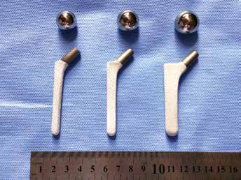
The prosthesis used in our study.
The Canine Femoral Head Replacement
In pre-operative preparation, the dogs were banned food and water for 12 hours. After the completion of intravenous anesthesia, anteroposterior and lateral pelvic radiographs were taken and the operation area was shaved. Before the operation, the cefmenoxime (50 mg/kg) would be used for intravenous drip. The animals were placed on the operating table on their left side, and the operation area was decontaminated by povidone-iodine, and covered with asepsis drapes. The hip joint was exposed through an anterior-lateral approach using standard sterile techniques. After an articular capsulotomy and section of the ligamentum teres, the femoral head was dislocated and resected by use of wire saws. After the femoral medullary cavity was curetted and rasped, the stem was implanted to the optimal position. After these procedures, the wound was thoroughly washed and closed in layers (Fig. 2). The postoperative anteroposterior and lateral pelvic radiographs showed that the prostheses were securely fixed and well positioned (Fig. 3) (21).
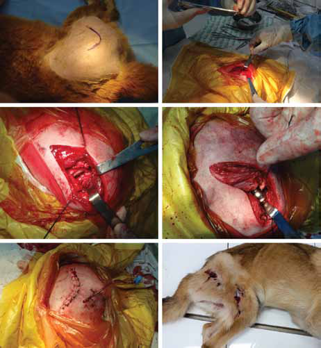
The process of building postoperative periprosthetic infection canine models.
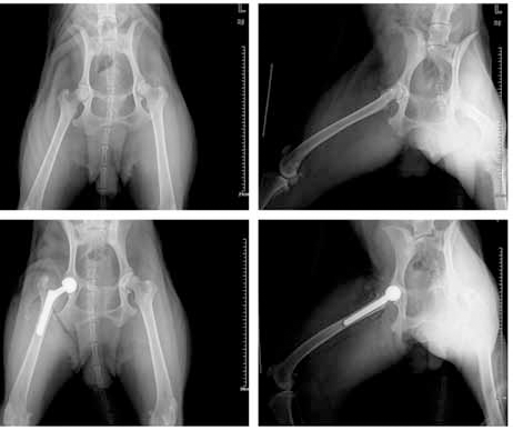
The anteroposterior and lateral pelvic radiographs of pre-operative and postoperative canine models.
The Enzyme-linked Immunosorbent Method
Canine RANKL ELISA Kit was purchased from LanpaiBIO (LanpaiBIO Co. Ltd., Shanghai, China). The content of RANKL was determined according to the manufacturer's protocol. The kit assay Canine RANKL level in the sample uses purified antibody to coat micro-titer plate wells to make solid-phase antibody. Samples, which include standards of known and unknown RANKL concentrations, are pipetted into coated microtiter wells. After incubating biotinylated anti-IgG is added, and combined streptavidin-HRP, the samples become antibody antigenenzyme-antibody complex, after washing completely, they are measured the optical density (OD) at 450 nm with microtiter plate reader to calculate Canine RANKL concentration by standard curve.
The kit assay Canine osteoprotegerin (OPG) level in the sample uses purified antibody to coat microtiter plate wells to make solid-phase antibody. Samples which include standards of known OPG concentrations and unknowns are pipetted into coated microtiter wells, after incubating, add biotinylated anti-IgG, and combined streptavidin-HRP, become antibody-antigen-enzyme-antibody complex, after washing completely, measure the optical density (OD) at 450 nm with microtiter plate reader, calculate Canine OPG concentration by standard curve. All two kits were performed at room temperature and data were relative to the control group.
Data Analysis and Statistic
The experimental data were represented by mean ± standard deviation (SD). One-way analysis of variance from the SPSS (Chicago, IL) 17.0 statistical software package was used to analyze the data. Results were considered to be significant at the 5% critical level (p<0.05).
Results
All surgeries were completed successfully. One of the dogs contracted infection on the fifth day after surgery and another dog in the experimental group was dead after 10 days of the bacteria fluid intra-articular injection. These two dogs were excluded from the analysis. And there was no animal death or dislocation of the prosthesis in any of the other animals. Although they had mild lameness immediately after surgery, the gait gradually returned to normal after one week. According to the observation of the animal models in the process of our study, we found that the sioux were developed around the operation incisions in all of the test group animals after 2 weeks of the bacteria fluid intra-articular injection. The prosthetic loosened in the proximal portion of right femur in the test group animals, based on our observation.
To analyze the results of RANKL and OPG content in the synovial fluid samples assayed by the ELISA method, we find that the ratio of RANKL/OPG showed a significant upward trend (p≤0.05) with time in the test group but the ratio of RANKL/OPG in the control group changed slowly over time (p>0.05). The ratio of RANKL/OPG value between the test and control group showed a significant upward trend, but had no statistical difference (p>0.05) over time (Fig. 4).
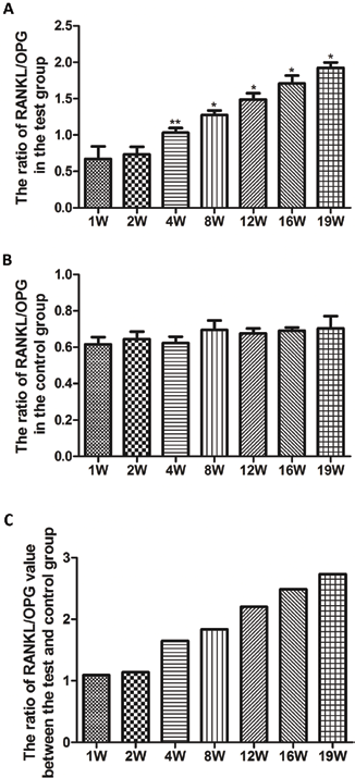
The expression of RANKL and OPG in the synovial fluid assayed by ELISA. (A) The ratio of RANKL/OPG in the test group. (B) The ratio of RANKL/OPG in the control group. (C) The ratio of RANKL/OPG value between the test and control group. (*p<0.05).
Discussion
The receptor activator of nuclear factor kappa B (RANK), its ligand (RANKL) and osteoprotegerin (OPG) as the final effectors of bone resorption have transformed our understanding of metabolic bone diseases and revealed novel therapeutic targets. Activation of the RANKL/RANK/OPG system is directly responsible for dramatic focal erosions that are observed in inflammatory arthritis and aseptic loosening of orthopedic implants (10–12, 22). To the best of my knowledge and belief, the signaling pathway of RANKL/RANK/OPG system has been widely discussed in osteoporosis, fracture healing, prosthesis loosening after joint replacement, and many other areas, while the prosthesis-related infection is a serious complication for patients after orthopedic joint replacement, which is currently difficult to treat and will prolong the time of hospitalization, but the mechanism of periprosthetic infection osteolysis has been little studied. In our clinical practice, we learn that the problem of bone defects in revision surgery infection is a common and thorny issue. Therefore, it is imperative to learn the pathogenesis of periprosthetic infection osteolysis. In our experiment, the result of the ELISA method for testing the expression of RANKL and OPG showed significant statistical difference (p≤0.05) in the test group. Furthermore, the ratio of RANKL/OPG was increased more obviously than the relative level of RANKL that was well matched in previous investigations about the RANKL/RANK/OPG system in osteolysis (20).
The canine model used in our experiment is a mature and classic model for the study of human hip arthroplasty. We gave the 5. epidermidis fluid (1×10 ∼8CFU) for intra-articular injection after the canine femoral head replacement and studied it for 19 weeks. Although this method has rarely been reported in the literature, we have integrated the previous studies in animal models of hip arthroplasty and periprosthetic joint infection experiments, so that the final decision on the application of this method has adequate theoretical basis. The arthroplasty dislocation rate after surgery, the surgical infection rate, mortality rate, and the success rate of infection model constructed in our study show that our experimental approach achieved the desired experimental target. There are some limitations in our study. First, the number of canine models was limited. In the experimental sample, two animals were not included in the experimental results, because one canine developed an incision infection after joint replacement surgery. In order to ensure that the experimental group and control group conditions were consistent before the bacterial injection treatment, it was excluded. The other dog was ruled out due to death after the bacterial injection. Second, we lacked the analysis of RANKL/RANK/OPG system on the genetic level. After the infection of the experimental animal we did not process bacteria culture because we considered the main purpose of this experiment was to study the mechanism of infection of periprosthetic osteolysis for the types of bacteria that cause infection of periprosthetic osteolysis. Maybe we need to explore further in the future. In this experiment, we did not collect and culture synovial fluid samples. If the joint fluid specimens were cultured and analyzed, the results of culture data and the relationship between the RANK system and the data were further analyzed. The results can be used as the next step of experimental research. Third, the content of RANKL in synovial fluid was extremely low. In future studies, the tissue specimens should be investigated. Despite these deficiencies, and according to the previous literature reports of experimental studies and our research, it seems reasonable to assume that the RANKL/RANK/OPG system is correlated with the incidence of periprosthetic osteolysis with septic loosening. Consequently, imbalance of RANKL/RANK/OPG system was related to periprosthetic osteolysis with septic loosening. We consider that osteolysis around the joint prosthesis results in a loosening of the prosthetic loosening system may be involved in an important system that plays a vital role in the aseptic or bacterial loosening process, and has no specificity. From the experimental results, we did not consider the RANK system as a diagnostic indicator of infection around the prosthesis, since the RANK system has an upward trend over time in the presence of bacterial loosening, but the difference is not statistically significant. After the animal model construction appeared periprosthetic osteolysis, if we choose a method such as MICRO-CT, further validation is also possible. Data in the literature have confirmed the RANK system as playing an important role in osteolysis. So, after we considered the fact that the RANK system was involved in osteolysis, no validation experiment included. In future, this method can provide a theoretical basis for the study of periprosthetic infection animal model. Besides, the RANKL/RANK/OPG system should be further studied and many other pathophysiological mechanisms of periprosthetic infection osteolysis should be explored to broaden the research direction of osteolysis.
Conclusions
The conclusion is that the RANKL/RANK/OPG system is correlated with the incidence of periprosthetic osteolysis with septic loosening. Consequently, RANKL/RANK/ OPG system imbalance was related to periprosthetic osteolysis with septic loosening.
Footnotes
Financial support: This work was supported by funding from the National Natural Science Foundation of China (No. 81272016).
Conflict of interest: The authors have declared no conflict of interest.