
Abstract
Patients with functional gastrointestinal disorders express antibodies against gonadotropin-releasing hormone (GnRH) in serum. One common cause of posterior laryngitis (PL) is extra-esophageal reflux, but a functional etiology has also been suggested. The aim of this study was to scrutinize patients with PL with regard to the presence of GnRH antibodies and to examine the association between antibodies and symptoms and reflux. Consecutive PL patients were included after examination. Serum was analyzed for the presence of antibodies using an enzyme-linked immunosorbent assay (ELISA) method and expressed as relative units (RU). Two age- and gender-matched healthy subjects per case served as controls. The prevalence of IgM GnRH antibodies in patients was 35% compared with 28% in controls (P = 0.06), with higher levels in patients (0.8 (0.3–2.2) RU) than in controls (0.2 (0.1–0.6) RU) (P = 0.007). The corresponding IgG antibody prevalences were 43% and 4%, respectively (P = 0.001), with no difference in levels (P = 0.70). There was no association between antibodies and clinical findings.
Introduction
Reflux of stomach contents into the esophagus is the most common cause of severe symptoms from the upper gastrointestinal tract. Although the exact mechanism is not known, reflux is associated with esophageal dysmotility in 40% to 50% of cases and reduced pressure of the lower esophageal sphincter (LES).1–3 Reflux leads to heartburn, acid regurgitation, and esophagitis,2,3 but reflux may also lead to more proximal signs such as posterior laryngitis (PL). PL is characterized by an inflammatory response of the posterior part of the glottic region causing symptoms such as chronic cough, hoarseness, globus, excessive throat clearing, voice fatigue, and throat pain. 4 Posterior laryngitis has been assumed to often depend on acid reflux, why proton pump inhibitors (PPI) is the choice of treatment.5,6 However, PL may be caused also by other mechanisms, and similar findings of the glottic region as in PL are described in healthy individuals without the typical symptoms of PL.7,8
Gonadotropin-releasing hormone (GnRH) is best known to bind to specific receptors on the pituitary, controlling the secretion of the sex hormones.9,10 Its presence and function as a neurotransmitter in the enteric nervous system (ENS) has recently been described. 11 Antibodies against GnRH have been observed in patients with primary functional bowel disorders such as irritable bowel syndrome (IBS), in patients with functional bowel symptoms secondary to primary Sjögren's syndrome, as well as in patients affected by unclear dysmotility disorders. Patients with organic gastrointestinal diseases such as inflammatory bowel disease, celiac disease, and scleroderma do not express antibodies.12,13 This raises the hypothesis that patients with different functional disorders, which often are associated and show an overrepresentation in women,14,15 may express GnRH antibodies as a common feature. We have recently shown how the majority of patients suffering from PL are women who are not improved by PPI treatment and express low health-related quality of life. 16 This is in accordance with the hypothesis of a functional etiology of proximal gastrointestinal and extra-esophageal complaints.8,17
The presence of GnRH antibodies in patients suffering from PL has not been investigated. The aim of the present study was to scrutinize consecutive patients with PL for the prevalence of acid reflux and the presence of GnRH antibodies and to examine the association between antibodies and other measured parameters.
Materials and Methods
This study was performed according to the Helsinki Declaration and was approved by the Regional Ethics Review Board of Lund University. All subjects gave written informed consent before entering the study.
Subjects and Methods
Consecutive patients over 18 years of age from the Division of Otorhinolaryngology, Skåne University Hospital, Malmö, Sweden, were invited from June 1, 2007 through May 31, 2011, to participate in the study if the diagnosis PL was made by fiber laryngoscopy. The diagnosis criteria for PL and, thereby, the criteria for inclusion in the study were thickening and/or edema of the posterior part of the glottic region in combination with one or several of the following symptoms: globus, hoarseness, excessive throat clearing, excessive phlegm, acid regurgitation/reflux, heartburn, coughing, voice fatigue, breathing difficulties. or a feeling of cramp in the throat. These symptoms were chosen as they are the most frequently described symptoms in this group of patients. 4 When seeing the ear, nose, and throat specialist, the patients were asked about the symptoms mentioned above, and perceived symptoms were registered in their medical records. In addition, information such as age, duration of symptoms, drug treatments, concomitant diseases, and body mass index (BMI) were registered. All patients were referred for 24-hour, single probe, pH monitoring in the proximal part of the esophagus and esophagogastroduodenoscopy (EGD) to examine the upper gastrointestinal tract. Blood samples for the analysis of anti-GnRH antibodies were collected at the time of the EGD. Exclusion criteria were pregnancy, mental illness, and serious illness such as severe heart, lung, liver, and kidney disease. Patients with Hepatitis B, Hepatitis C, or HIV/AIDS were not included.
Thirty-seven of the 60 patients invited agreed to give blood samples for the analysis of anti-GnRH antibodies and were included in the study. Five of these 37 patients could not tolerate the catheter used for 24-hour pH monitoring. All patients included tolerated the EGD.
Ambulatory 24-hour pH monitoring
All participants were instructed to discontinue proton pump inhibiting therapy seven days before monitoring and to discontinue other acid inhibitors 16 hours before monitoring. Participants were asked to avoid acidic beverages such as juice during the 24-hour period of pH monitoring and to fast 4 hours before the catheter was introduced. The positioning of the catheter was done in the Diagnostic Center of Imaging and Functional Medicine with the aid of fluoros-copy (Philips Multidiagnost Eleva, CA, USA). The catheter was introduced through the nose and under fluoroscopic control positioned in the proximal part of the esophagus, 5 cm below the upper esophageal sphincter.
Esophageal pH monitoring was performed using an antimony pH electrode with an internal reference electrode (Versaflex, Sierra Scientific Instruments, Los Angeles, CA, USA). Before each study, the pH-probe was calibrated in buffer solutions of pH 7 and pH 1. An episode of acid reflux was defined as a decrease in esophageal pH below 4 for more than 10 seconds. Previously established upper limits of normal acid exposure in clinical studies, with pH < 4 for 1% of total time, were used in the analysis of the data.18,19 The data were stored on a portable digital recorder (Digitrapper pH400, Synectics Medical, Stockholm, Sweden). Data were analyzed with the aid of commercially available software (Polygram NET, SynMed Medical, Stockholm, Sweden).
Measurement of human antibodies against gonadotropin-releasing hormone
Blood samples were drawn from patients and the serum was separated and kept frozen at −20 °C until analyzed. Analysis of anti-GnRH antibodies was carried out by an enzyme-linked immunosorbent assay (ELISA) method slightly modified on the basis of the results described in previous studies.11,12 The wells of microtiter plates were coated with human GnRH (L7134, Sigma, St Louis, MO, USA) for an overnight incubation at 4 °C, and, thereafter, the plastic wells were blocked with 0.5% fish gel solution (G7765, Sigma, St Louis, MO, USA) in phosphate buffered saline (PBS) containing 0.05% Tween-20 (PBS-T). Serial dilutions of patient serum (1/100, 1/500 and 1/2500 in PBS-T) were then added to the plates and incubated for 2 hours at room temperature and overnight at 4 °C. After rinsing with PBS-T, deposition of autoantibodies directed to GnRH was detected using biotinylated rabbit antihuman IgM (673211, MP Biomedicals, Solon, OH, USA) or IgG antibodies (ab7159, ABcam, Cambridge, MA, USA) appropriately diluted in PBS-T. After another incubation for 2 hours at room temperature, the plates were washed, and the bound, biotinylated antibodies detected by alkaline phosphatase-conjugated streptavidin (405211, Biolegend, San Diego, CA, USA) and incubated for 1 hour at room temperature. To develop a color reaction, a phosphatase substrate kit (37620, Pierce, Rockford, Ill, USA) was used. The absorbance at 405 nm was measured after 2 hours of incubation at room temperature. A plasma pool from healthy blood donors was included on each ELISA-plate for measurements of the variation. The plasma pool was used for the calculation of the intraassay and interassay coefficient of variations, which were 11.5% and 16.1%, respectively, for IgM and 11.5% and 25.4%, respectively, for IgG. Antibody levels are presented as relative units (RU) (absorbance values after subtraction of background levels and multiplied by 100). Relative units over 0 were considered as a positive antibody level. 13 The controls were chosen from a cohort of healthy blood donors previously described in detail. 13 Over a period of five months (October 1996–February 1997), blood donors were offered antibody screening for gastrointestinal diseases. To be able to include all blood donors in Malmö, sera from male donors were collected over a 3-month period and from female donors over a 4-month period (in accordance with their regular donation intervals). A total of 1970 donors were included. During this period, 2135 blood donations took place, which means that at least 92% of donors agreed to be included. From this sample cohort, 50 men and 50 women from each 10-year age span period, between 20 and 70 years, were randomly included. As few blood donors are over the age of 60 years, only 16 women and 40 men were included in the age group 60 to 70 years. In total, 456 controls were examined during the same time period as the patients. From this cohort, two age- and gender-matched controls were randomly extracted for each patient in this study.
Statistical analyses
The data were analyzed using the statistical software package SPSS for Windows, release 19.0 (IBM Corporation, Armonk, NY, USA). Values are expressed as median and interquartile range (IQR). Group-wise differences were tested by using the Mann Whitney U test or Fisher exact test. Correlations were calculated by the Spearman test. Results where P < 0.05 were considered statistically significant.
Results
Patient characteristics
Thirty-seven patients with verified PL (20 women) with a mean age of 56 (range 40 to 69) years were included (Table 1). Esophagogastroduodenoscopy found no ulcerations or tumors, but 7 patients suffered from esophagitis, 10, from Barrett's esophagus, 13, from hiatal hernia, and 12 (of 32 examined), from proximal esophageal reflux. The most common symptoms present were globus (65%), excessive phlegm (46%), and hoarseness (32%) (Table 2). Apart from symptoms associated with PL, 7 patients also described dysphagia.
Characteristics of patients with posterior laryngitis.
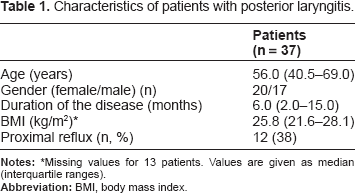
Missing values for 13 patients. Values are given as median (interquartile ranges).
The prevalence of various symptoms in patients with posterior laryngitis (n = 37).
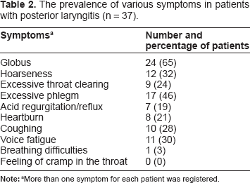
More than one symptom for each patient was registered.
Antibodies against gonadotropin-releasing hormone
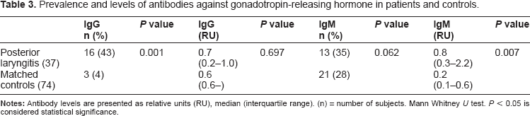
The prevalence of IgM antibodies against GnRH in patients was 35% compared with 28% in controls (P = 0.06), and the antibody level was significantly higher in the patients (P = 0.007) (Fig. 1A and Table 3). The prevalence of IgG was 43% in patients and 4% in the controls (P = 0.001), with no difference in the level of antibodies between the patients and controls (P = 0.70) (Fig. 1B and Table 3). There was no association between the expression of IgM and IgG antibodies (P = 0.79), but one or both of these antibodies were found in 24 of 37 (65%) patients compared with 24 of 74 (32%) controls (P = 0.002). Neither was there any association between the presence of antibodies and symptoms, duration of symptoms, esophageal diseases or BMI (data not shown). The level of antibody titer did not differ between those who had symptoms and those who did not. There was no correlation between number of symptoms and presence or levels of antibodies (data not shown).
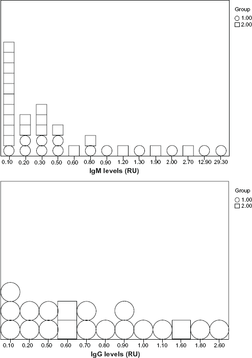
The level of IgM and IgG antibodies expressed as relative units (RU). (
Prevalence and levels of antibodies against gonadotropin-releasing hormone in patients and controls.
Discussion
The present study showed that patients with PL had few organic findings on 24-hours pH monitoring and EGD examination. Thirty-eight percent had pathological proximal acid reflux, and 46% had signs of distal acid reflux. The majority, 65%, expressed antibodies against GnRH in serum compared with 32% in controls.
Gonadotropin-releasing hormone is secreted by the hypothalamus, and its most important effect is on the pituitary, stimulating gonadotropin synthesis and secretion. It is a crucial neuropeptide in reproductive physiology and sexual behaviour. 9 Peripherally, GnRH and GnRH receptors have been found in the rat myenteric plexus and the intestinal epithelium.20,21 We have recently described the expression of GnRH in human myenteric neurons. 11 The effect on the ENS is not completely evaluated, but GnRH has been shown to inhibit the release of gastric secretion and gastrin release in dogs, 22 to stimulate motor function in the gastrointestinal tract in female rats, 23 and to restore motor function in a patient suffering from chronic intestinal pseudo-obstruction. 24 Although it has now been described in several studies that patients with IBS and dysmotility express GnRH antibodies,11–13 the effects of GnRH and/or its antibodies on the normal physiology of the gastrointestinal tract, as well as on pathological processes and symptom development, remain to be determined. We do not know whether there is a difference between the expression of IgM and IgG antibodies in these patients. Some chronic inflammatory diseases present themselves with IgM antibodies instead of IgG antibodies. Probably, the expression of IgM antibodies is long-standing in this entity, as the patients had been sick for several years before inclusion in the study.
Many factors, such as rising obesity rates, greater consumption of medications affecting esophageal function, and potentially changing prevalence rates of Helicobacter pylori infection, have been discussed as the etiology of reflux. 25 Although PL is considered to depend on acid reflux, 4 only 12 of 32 (38%) patients had a pathological 24-hour pH monitoring as proof of proximal acid reflux, and 17 of 37 (46%) showed signs of distal acid reflux. Furthermore, we have recently shown that 63% of patients with PL still have symptoms after PPI treatment, 16 showing the need to consider other etiologies than acid reflux as a cause of PL. Factors such as pepsin, bile, infections, allergy, and smoking have been evaluated and discussed.7,8 Nobody has examined whether signs of functional disorders are present in this patient group, despite that the disease is characterized by female predominance as in other functional disorders.4,14,15 The hypothesis that the symptoms of PL might partly be functional has been raised.8,17 This led us to perform the present study. Accordingly, 65% of patients with PL expressed either IgM or IgG antibodies in serum and had higher antibody levels than controls, as found in functional gastrointestinal disorders previously.12,13 No association between signs of reflux at EGD or pH monitoring and antibodies was found. Neither was there any association between symptoms and antibody level. It is a well-known phenomenon that there is a weak or absent correlation between objective signs and subjective symptoms from the gastrointestinal tract. The reason is unclear, but may depend on different central processing of visceral, afferent information in healthy controls and patients suffering from functional bowel diseases. 26 Psychological factors have a great impact in the pathophysiology of visceral hyperalgesi.27,28 The presence of antibodies observed in this study strengthens the hypothesis that PL may be a functional disorder in a subgroup of patients presenting themselves with laryngitic inflammation and symptoms, as GnRH antibodies are described in patients with functional disorders without any association between antibodies and symptoms and signs.11–13 Gonadotropin-releasing hormone and its receptors are present both centrally and peripherally, and deregulation of this peptide and/or its receptor may be involved in the pathophysiology of functional disorders.
The controls used were healthy blood donors. However, functional disorders are not exclusion criteria for blood donors and are very common in the general population,14,15 and the controls were not asked about symptoms. Thus, among the controls may be hidden some persons with functional disorders as well, explaining some of the antibodies in this group. If only persons who had denied all types of functional disorders had been used as controls, the differences between controls and patients might have been greater.
One of the limitations of this study is the small sample size. However, it is necessary to perform small pilot studies before testing our hypothesis in larger cohorts of hundreds of patients. In future studies, it seems that it would be more relevant to focus on functional etiologies to PL and not only focus on acid reflux, which seems to be a lesser problem among these patients. Another important task is to elucidate whether the presence of antibodies is a primary or a secondary phenomenon, which may have an impact on associations. The test method also needs to be further evaluated in relation to the optimal cutoff level. In our ELISA, we considered all relative antibody units above 0 as positive expression. If a higher baseline level of antibodies present in serum had been regarded as positive expression, more controls than patients would have been below this level. When more patients have been studied, the cutoff level for positive expression should be redefined.
Conclusions
Patients with PL express IgG antibodies against GnRH in serum in higher prevalence, whereas the level of IgM antibodies is increased compared to controls. Only one-third of the patients had objective signs of proximal acid reflux in the esophagus. No associations between antibody expression and other parameters were found. A subgroup of patients with PL may suffer from functional disease, when no other etiology is found.
Funding
This study was sponsored by grants from the Crafoord and Bengt Ihre Foundations and from the Development Foundation of Region Skåne.
Competing Interests
Author(s) disclose no potential conflicts of interest.
Author Contributions
All authors participated in the design of the study. Collected the blood samples and data from the Division of Otorhinolaryngology: HP. Performed the ELISA analyses: RA and GNF. Contributed to the statistical analyses and wrote the manuscript: HP and BO. Supported the study financially (Crafoord- and Bengt Ihre Foundations, and Development Foundations of Region Skane): BO. All authors contributed to the manuscript with constructive criticism and read and approved the final manuscript.
Disclosures and Ethics
As a requirement of publication author(s) have provided to the publisher signed confirmation of compliance with legal and ethical obligations including but not limited to the following: authorship and contributorship, conflicts of interest, privacy and confidentiality and (where applicable) protection of human and animal research subjects. The authors have read and confirmed their agreement with the ICMJE authorship and conflict of interest criteria. The authors have also confirmed that this article is unique and not under consideration or published in any other publication, and that they have permission from rights holders to reproduce any copyrighted material. Any disclosures are made in this section. The external blind peer reviewers report no conflicts of interest.
Footnotes
Acknowledgements
We thank Ola Thorsson, Department of Clinical Sciences, Nuclear Medicine, Diagnostic Centre of Imaging and Functional Medicine, Skåne University Hospital, Malmö, Klas Sjöberg who collected samples from healthy blood donors, and Peter Höglund, Region Skånes Kompetens Centrum (RSKC) for assistance with the statistical calculations.