
Abstract
Aim
To study the role of Doppler imaging in prediction of high-risk pregnancies and their outcomes.
Methods and Material
This prospective study in a setup of tertiary-level care center includes 500 high-risk pregnant women from rural and urban sectors and evaluates the predictive values of various Doppler indices.
Results
Out of 500 patients, 110 patients had abnormal Doppler among them, 70 patients had abnormal uterine artery Doppler, and 50 patients had abnormal umbilical artery Doppler flow indices. In all, 10 patients had both umbilical artery and uterine artery abnormal Doppler indices. When uterine artery was abnormal (70 patients), 20 patients had preeclampsia, 10 patients had pregnancy-induced hypertension (PIH), and 25 patients had intrauterine growth restriction (IUGR). Systolic/diastolic (S/D) ratio and notch had sensitivity of 60% and positive predictive value of 33.3% and 37.5%, respectively. When umbilical artery was abnormal (50 patients), 10 had preeclampsia, 15 had PIH, and 15 had IUGR. S/D ratio had the highest positive predictive value of 40%; sensitivity is same for all. In uterine artery, combination of parameters had the best sensitivity of 80%, followed by notch and S/D ratio. In umbilical artery, combination of parameters, S/D ratio, and RI (resistance index) had sensitivity of 40%; specificity of all the indices was 91-96%. In all, 20 patients had bilateral notch, and among them 15 developed preeclampsia and 15 developed IUGR. When both uterine and umbilical artery Doppler were abnormal (10 patients), all patients had preeclampsia and IUGR.
Conclusion
Therefore, Doppler study may be used for the prediction of preeclampsia and IUGR to reduce the maternal and perinatal morbidity and mortality.
Introduction
Despite recent advances in antenatal care, preeclampsia remains the major cause of maternal and perinatal morbidity and mortality. So early screening of preeclampsia and fetal growth restriction may allow vigilant antenatal surveillance and appropriate timing of fetal delivery in order to avoid serious sequelae.
Doppler techniques have been the focus of interest and research activity in obstetrics since the initial report of signals from the umbilical artery by Fitzgerald and Drumm. 1 The first application of Doppler velocimetry in obstetrics was reported by Fitzgerald and Drumm 1 and McCallum et al. 2 It has been long assumed that insufficient uterine, placental, and fetal circulations result in adverse pregnancy outcomes and that those abnormalities can be defined by the use of Doppler ultrasonography. 3
Uteroplacental blood flow decreases in pregnancies that are complicated by hypertension and intrauterine growth restrictions (IUGRs). This decrease is associated with a pathologic condition of spiral arteries, thought to arise during placentation in the first trimester of pregnancy; thus, it might be possible to predict the development of these conditions by assessing uteroplacental blood flow early in pregnancy with color Doppler. 4
The result is abnormal uteroplacental blood flow, and this has led to the idea of using Doppler assessment of uterine and umbilical arteries velocity waveforms as a method of screening for these antenatal complications. An abnormal test result is represented by an abnormal flow velocity ratio (systolic/diastolic (S/D) ratio), resistant index, or the presence of an early diastolic notch.5,6
Despite the absence of any treatment that reverses the disease process once started, screening for preeclampsia and IUGR has been a major clinical and research issue since the disease was first reported. So we have planned to study the role of Doppler imaging in prediction of high-risk pregnancies and their outcomes.
Materials and Methods
A total of 500 women with high-risk pregnancy attending antenatal clinic at Panna Dhai Rajkiya Mahila Chikitsalaya, Udaipur, were subjected to uterine and umbilical artery Doppler study. Women with high-risk pregnancy between 26 and 30 weeks of gestation were studied in this prospective study over a period of two years from May 2009 to May 2011 with color Doppler. Umbilical and uterine artery flow velocity forms were studied. In both uterine and umbilical artery, S/D ratio and RI (resistance index) were studied. S/D ratio and RI of >95th percentile from standard values were classified as abnormal. In uterine artery presence and absence of early diastolic notch was noted. In umbilical artery, absent and reversed diastolic flows were studied. Informed consent was obtained from the women who underwent color Doppler study. The outcomes of the pregnancies were evaluated by review of the hospital records after delivery. This study was approved by institutional research board (IRB) of the institute.
The accurate gestational age (GA) was estimated by the last menstrual period (LMP) with 28-30 days regular cycles. When a woman did not fulfiled this criteria, accurate expected date of delivery (EDD) was calculated by ultrasound examination before 21 weeks of GA.
Inclusion criteria
History of preeclampsia or eclampsia in previous pregnancy Pre-existing medical disorders like:
Diabetes,
Renal disease,
Epilepsy,
Autoimmune disease,
Thrombophilia, and
Hypertension
History of IUGR or still birth.
History of abruptio placentae.
Preeclampsia or pregnancy-induced hypertension (PIH) current.
Nulliparity.
Extremes of age (<20 years and >35 years).
Exclusion criteria
Patient with congenital anomaly of fetus, multiple gestations.
Patients those who are not getting booked for delivery at Panna Dhai Rajkiya Mahila Chikitsalaya in Udaipur.
Patients with unreliable LMP details and not confirmed by early ultrasound.
These patients were followed up till delivery, and details of pregnancy events, labor and delivery, and neonatal outcome were noted. The abnormal pregnancy outcomes considered were PIH and IUGR. Abnormal perinatal outcomes considered were intrauterine death, five minutes Apgar score, Neonatal intensive c are unit (NICU) admission, and birth weight. This research complied with the principles of the Declaration of Helsinki.
Statistical analysis
All the data were entered in Microsoft excel sheet. The data were analyzed using SPSS version 16 for Windows. The sensitivity, specificity, and predictive value were calculated.
Results
In the study period, we enrolled 500 pregnant females who fulfilled the inclusion criteria. In our study, 11% mothers were of age less than 20 years, 55% (maximum) patients were in the age group of 21-25 years, 33% were between the age group of 26-30 years, and 1% (minimum) were of age >31 years. Most patients were primi gravida and second gravida. A total of 38% of the patients were educated up to the higher secondary and 17% were graduate. In all, 58% of patients had vaginal delivery and 30% of patients had emergency LSCS and 12% of patients had elective lower segment cesarean section (LSCS).
Preeclampsia and Doppler imaging
S/D ratio and notch on uterine artery Doppler imaging individually had sensitivity of 60% and positive predictive values of 33.3% and 37.5%, respectively, for preeclampsia. Notch as a single parameter was the best indicator with the highest sensitivity and positive predictive value. However, combination of both parameters was the best indicator for predicting preeclampsia with sensitivity and specificity of 80% and 89.4%, respectively (Table 1).
Uterine artery Doppler in predicting preeclampsia.

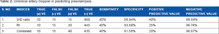
S/D ratio had the highest positive predictive value (40%) when compared to RI, and they were combined for predicting preeclampsia on umbilical artery Doppler imaging. On umbilical artery, Doppler combination of parameters (S/D ratio and RI) had sensitivity of 40%, and specificity of all the indices was 91%-96% (Table 2).
Umbilical artery Doppler in predicting preeclampsia.
PIH and Doppler imaging
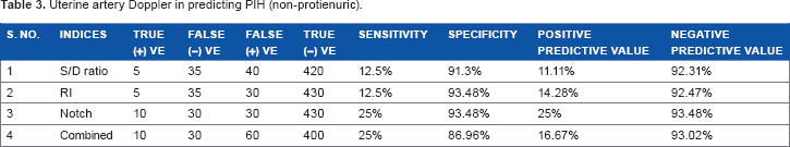
Notch on uterine artery Doppler imaging was the best indicator with the highest sensitivity (25%) and positive predictive value (25%) for PIH as compared to other indices (Tables 3 and 4).
Uterine artery Doppler in predicting PIH (non-protienuric).
Umbilical artery Doppler in predicting PIH (non-proteinuric).

IUGR and Doppler imaging
Notch on uterine artery Doppler as a single parameter was the best indicator with the highest sensitivity and positive predictive value of 50% for IUGR. However, combination of parameters had the best sensitivity of 62.5%.
On umbilical artery, Doppler S/D ratio had the best positive predictive value (40%) and RI had the best sensitivity (42.86%) for predicting IUGR (Tables 5 and 6).
Uterine artery Doppler in predicting IUGR.

Umbilical artery Doppler in predicting IUGR.

When there was a notch in uterine artery Doppler imaging, 37.5% of patients developed preeclampsia, 25% of patients developed PIH, 50% of patients developed IUGR, 37.5% of patients had intrauterine death, and 12.5% of patients had still birth. This indicates that notch in the uterine artery was associated with poor pregnancy outcome.
When umbilical artery Doppler S/D ratio was abnormal, 40% of patients developed preeclampsia, 20% of patients developed PIH, and 40% of patients developed IUGR. Five patients had absent end diastolic flow, which was associated with preeclampsia, IUGR, and intrauterine death. This indicates that absent end diastolic flow was associated with poor pregnancy outcome.
When both uterine and umbilical Doppler were abnormal, 100% patients had developed preeclampsia and IUGR. When both normal, only 1.28% patients developed preeclampsia, 3.85% developed PIH, and 2.56% patients developed IUGR.
Discussion
In this prospective study in a setup of tertiary level care centre, which includes women from rural and urban sectors, the predictive values of various Doppler indices have been evaluated.
In our study, the prevalence of preeclampsia was 5%, which was similar to that quoted by Bewley et al 7 and Irion et al, 8 which was 4.6% and 4%, respectively. Prevalence of SGA less than 10 percentile (IUGR) was 8%, which was similar to that quoted by North et al (6.6%). 4
Out of 500 patients, 110 patients had abnormal Doppler; among them, 70 patients had abnormal uterine artery Doppler and 50 patients had abnormal umbilical artery Doppler flow indices. In all, 10 patients had both umbilical artery and uterine artery abnormal Doppler indices.
Out of 70 patients who had abnormal uterine artery, 45 had abnormal S/D ratio and 35 had abnormal RI. In all, 40 patients had persistent early diastolic notch; among them, 20 patients had bilateral notch and 20 patients had unilateral notch. There were 50 patients with abnormal umbilical Doppler; among them, 25 had abnormal S/D ratio, 40 had abnormal RI, and 5 had absent end diastolic flow.
Out of these 110 patients, 16 developed preeclampsia with a sensitivity of 60%, 40%, and 60% for uterine artery S/D ratio, RI, and notch, respectively. The specificity was 93%-94% for all the indices. This is similar to the result obtained by Kurdi et al. 9 Bhattacharya et al 10 did study in 179 women and found that sensitivity and specificity of abnormal uterine artery Doppler for prediction of preeclampsia were 73.33% and 86.48% in high-risk pregnancies. The positive predictive value was 33.3%, 28.6%, and 37.5% for S/D ratio, RI, and notch, respectively. This indicates that notch is a better predictor of preeclampsia. This is similar to opinions by Bower et al 11 and Antsaklis et al. 12
Out of 50 patients with abnormal umbilical artery, 10 patients developed preeclampsia with a sensitivity of 40%, 40%, and 100% for S/D ratio, RI, and absent end diastolic flow, respectively, and with a specificity of 96.84%, 93.68%, and 100% for S/D ratio, RI, and absent end diastolic flow. Mirza et al 13 did study in 268 women, and there was 57 cases with abnormal Doppler. Of these, preeclampsia was diagnosed in 14% cases. Positive predictive values of S/D ratio, RI, and absent end diastolic flow was 40%, 25% and 100%, respectively. This indicates that umbilical artery Doppler is more predictive than uterine artery Doppler.
In all, 40 patients had IUGR babies, which was predicted by abnormal uterine artery Doppler in 25 cases with a sensitivity of 37.5%, 25% and 50% for S/D ratio, RI, and notch, respectively. It is similar to opinion by Irion et al, 8 North et al, 4 and Bower et al. 11 The specificity was 93.48%, 94.56%, and 95.65% for S/D, RI, and notch respectively. Velauthar et al 14 did meta-analysis (18 studies of 55,974 women) and found that the sensitivities of abnormal uterine artery Doppler for predicting preeclampsia and fetal growth restriction were 26.4% and 15.4%, respectively, and specificities were 93.4% and 93.3%, respectively. The positive predictive value of notch was 50%, which is the highest. Here again notch was the best predictor than any other indices.
Out of 50 patients with abnormal umbilical artery Doppler, 15 patients had IUGR births with a sensitivity of 25% and 42.86% for S/D ratio and RI, respectively. It is similar to opinion by Antsaklis et al 11 and Beattie et al. 15 Romero et al 16 studied 43 women, and among them, 52% had abnormal umbilical Doppler. They found that abnormal umbilical Doppler is associated with lower birth weight, lower Apgar score, and significant neonatal morbidity. Sensitivity of absent end diastolic flow was 100%.
Ten patients had abnormal Doppler in both uterine and umbilical arteries. All patients developed preeclampsia and IUGR with a sensitivity and specificity of 100%.
In our study, 10 patients had abruptio placentae and intrauterine fetal death. All patients had abnormal Doppler indices in uterine artery. Out of 10 patients, 5 patients had bilateral notch in uterine artery and abnormal indices in both uterine and umbilical arteries and intrauterine fetal death at 35 weeks of gestation (IUGR). The same patients also developed preeclampsia. Other five patients had abnormal S/D ratio in uterine artery and unilateral notch and developed PIH and intrauterine fetal death at 33 weeks of gestation with an average birth weight of 1.8 kg.
There were five patients of preeclampsia who had bilateral notch in uterine artery, and they delivered after induction, an IUGR neonate at 28-29 weeks of gestation with a mean weight of 700 g; all were still born. There were five patients with absent end diastolic flow in umbilical artery and bilateral notch in uterine artery and had intrauterine death at 30-32 weeks of gestation with IUGR baby delivered by induction.
Results are similar to other studies (North et al, 4 Irion et al, 8 Bower et al, 10 and Bewley et al 7 ) except PPV for notch is more predictive as the present study was done from 26 weeks of gestation that reduces false-positive rate. This shows abnormal Doppler is associated with poor pregnancy outcome.
The antenatal interventions, which has been proved evidence based to reduce the incidence of IUGR, include antiplatelets for preeclampsia, balanced energy protein supplementation, multiple micronutrient supplementation, insecticide-treated bed nets, and intermittent preventive treatment of malaria in pregnancy.7,17,18
Conclusion
Combination of uterine and umbilical artery Doppler is the best indicator for prediction of preeclampsia and IUGR. Diastolic notch in the uterine artery as a single parameter is better than the individual Doppler indices in uterine artery. Absent end diastolic flow is the best predictor of preeclampsia and poor fetal outcome. Women with normal Doppler flow in both uterine and umbilical arteries constitute a group of low risk of developing obstetric complications compared to those with abnormal flows. Uterine and umbilical artery may be included in hospitals to identify a group of patients at risk of developing preeclampsia and poor fetal outcome. Therefore, Doppler study may be used for the prediction of preeclampsia and IUGR to reduce the maternal and perinatal morbidity and mortality.
Author Contributions
Conceived and designed the experiments: TN, DS. Analyzed the data: TN, DS, MC. Wrote the first draft of the manuscript: MC, DS, TN. Contributed to the writing of the manuscript: RPN, AP. Agree with manuscript results and conclusions: TN, DS, MC, RPN, AP, SK. Jointly developed the structure and arguments for the paper: TN, DS, MC, RPN, AP, SK. Made critical revisions and approved final version: TN, DS, MC, RPN, AP, SK. All authors reviewed and approved of the final manuscript.