
Abstract
Echocardiography accounts for nearly half of all cardiac imaging techniques. It is a widely available and adaptable tool, as well as being a cost-effective and mainly a non-invasive test. In addition, echocardiography provides extensive clinical data, which is related to the presence or advent of different modalities (tissue Doppler imaging, speckle tracking imaging, three-dimensional mode, contrast echo, etc.), different approaches (transesophageal, intravascular, etc.), and different applications (ie, heart failure/resynchronization studies, ischemia/stress echo, etc.). In view of this, it is essential to conform to criteria of appropriate use and to keep standards of competence. In this study, we sought to review and discuss clinical practice of echocardiography in light of the criteria of appropriate clinical use, also we present an insight into echocardiographic technical competence and quality improvement project.
Introduction
Echocardiography is increasingly used as a non-invasive diagnostic tool, as well as a method to monitor the progression of many cardiac conditions. The increased use of echocardiography is related to a certain extent to the new available modes (tissue Doppler imaging, strain imaging, speckle tracking imaging, three-dimensional mode, etc.), to the emerging indications (study of myocardial viability and resynchronization, etc.), as well as to the advent of new approaches (intracardiac echo, portable echo, etc.). This fact had a dramatic impact on echocardiography use and potential misuse. In view of that, we suggest that echocardiography must be achievable within a consistent existing framework of clinical standards in current cardiological practice, in order to yield a better clinical outcome, to prevent potential misuse and to preserve resources.
Method
Through a MEDLINE/Pubmed research, we used the terms “echocardiography; appropriate use”, “echocardiography; competence” and “echocardiography; recommendations”. The search started from 1985, selected articles mainly address the clinical outcome along with the managerial aspect of echocardiography rather than its pure technical feature. Moreover, the terms “clinical benefit; training; competence; appropriate use; quality improvement; sonographer behavior” were specifically searched in titles, abstracts, keywords and conclusions and subsequently were used as additional but not exclusive relevance criteria. One-hundred-and-thirty-one articles were analyzed and only 34 articles found to be relevant were selected; among these, there were 17 review papers, 13 original researches, 2 cases reports and 2 editorials.
Appropriate Clinical Use of Echocardiography
The continuous expansion of clinical applications in echocardiography is related to many factors including its non-invasive nature and cost-effectiveness, along with a significant recent technical improvement. However, this fact should not dissuade from the appropriate use of criteria for judicious implementation of echocardiography. In his 2011 report, 1 The American College of Cardiology Foundation (ACCF), in partnership with many key specialty and subspecialty societies, examined the suitability of transesophageal and transthoracic echocardiography (TTE) in different clinical conditions. Two-hundred-and-two criteria were analyzed and indications were classified as appropriate when the results of TTE were expected to change patient management and improve clinical outcome. The authors used a model of scoring based on an independent technical panel with a scale of 1 to 9: a score of [7-9] designates appropriate use, [4-6] designates uncertain use, and [1-3] designates inappropriate use. Among the 202 criteria, 97 were classified as appropriate, 34 as uncertain, and 71 as inappropriate. Of note, the ACCF document 1 states that “appropriate use of criteria for echocardiography has the potential to impact physician decision making, healthcare delivery, and reimbursement policy”.
Patil et al. 2 evaluated the appropriate use of criteria (AUC) of TTE according to the ACCF document 1 in a series of 1,825 consecutive patients who underwent TTE for clinical reasons. The authors found that TTE use was appropriate in 82% of cases, inappropriate in 12.3%, and uncertain or unclassifiable in 5.7%. Similarly, Matulevicius et al. 3 performed a retrospective review of electronic medical records by two independent cardiologists and found that 91.8% of the TTE use was appropriate. However, only one third of the appropriate tests resulted in active change in care and the authors evoked the need for a better implementation of ACCF criteria 1 to improve clinical outcome. Likewise, Armstrong et al. 4 questioned whether “No Change in care is equivalent to No Benefit?” and suggested that education of physicians may be required in order to achieve a more efficient use of the AUC according to the ACCF document. 1
In view of that, we consider that the AUC of the ACCF 1 provides a framework to guide general cardiologists, clinicians, and echocardiographists for the judicious use of echocardiography. Nevertheless, it does not imply a “perfect use” or an “optimal outcome” in real world.4,5 However, we estimate that this document is an outstanding report representing a cornerstone in current echocardiography practice that helps to restrict abuse, preserve resources, and improve clinical outcome.
Technical Competence
Competence in echocardiography is not only a technical component, it also requires appropriate knowledge, developed skills, and a positive attitude addressed to clinical benefit. It includes patient selection, study performance, interpretation and reporting, then trying to translate the whole process into better clinical outcome. The main mission statement of the European Association of Echocardiography (EAE) 6 is “to promote excellence in clinical diagnosis, research, technical development, and education in cardiovascular ultrasound in Europe”. We consider that the content of this statement should reflect the mission of every national echocardiography society worldwide in order to yield international echocardiographic standards implemented in every healthcare institution. However, in addition to these recommendations, we estimate that personal effort and awareness, together with local administrative regulations, are essential to ensure professional standards and ongoing competence in echocardiography.
Basic Training and Professional Standards
A basic training aims to acquire the fundamentals of ultrasound principles and sonoanatomy of the heart, including both theoretical and practical elements. Diagnostic medical ultrasonography in humans utilizes transducers of 1 to 20 MHz that generate ultrasound waves described in terms of frequency (Hz), wave length (mm), amplitude (decibels) and velocity (m/sec). TTE performance characteristically uses parasternal short and long axis together with apical views; it is typically useful for assessment of chambers dimensions, ventricular systolic and diastolic function, myocardial thickness, aortic root size, pulmonary artery pressure, pericardium and valvular morphology and function. In addition, sonographers may acquire specific expertise in echo approaches and applications (ie, transesophageal echocardiography, contrast echocardiography, pediatric echocardiography, echocardiography in congenital heart disease, stress echocardiography, intracardiac echocardiography). Moreover, according to the availability of equipment and competencies, operators may use advanced echocardiographic modalities (ie, three-dimensional mode, strain and speckle tracking imaging, tissue characterization, etc.).
Different stages of training and competence in diagnostic techniques are suggested.6,7 Stage (1) represents appropriate patient selection and study interpretation without performing the technique; stage (2) represents technical experience with mentored practice; and stage (3) represents independent and advanced practice with the ability to teach and coach others. Price et al 8 classified the levels of performance in echocardiography in intensive care as follows: Basic level, it represents focused echocardiographic expertise in life support (ie, tamponade, shock, etc.), and the operator (cardiologist or intensivist) may suggest referral to level 1, 2, 3; Level 1: performance of TTE, performance of all standard views, recognition when referral is indicated; Level 2: the operator accepts referral from level 1, performs comprehensive TTE and transesophageal echocardiography, evaluates “all” cardiovascular abnormalities, is involved in teaching and research; and Level 3: Echo specialist, with a post “exclusive” in echocardiography, highly skilled for standard and advanced techniques including “all” modalities and applications, involved in research, teaching and training, accepts referral from all levels.
Multiple training and accreditation programs are emerging in many countries. Some of these are tailored to local or national needs and aim to train clinicians for use of bedside ultrasound for the critically ill patients including fast assessment diagnostic echocardiography (FADE) 9 and focused echocardiography entry level (FEEL). 10 Manasia et al. 11 reported a significant clinical utility of goal-directed TTE performed by noncardiologist intensivists using portable echo machines in critically ill patients. Table 1 illustrates a schema of formative program in echocardiography as recommended by the American Society of Cardiology. 12
Formative program based on the Recommendations of the American Society of Cardiology.

Preserving professional standards is not only providing a good echocardiographic product, it is also an education and a culture targeting the clinical benefit and patient outcome. In view of that, we consider that sonographer's availability, appropriate patient selection, recognition of emergencies, and detection of the potential need for referral to an upper level are hallmarks of professionalism. In this perspective, we estimate that the time interval from ordering until achievement of the echocardiography study is critical and should be adapted to the clinical setting. Accordingly, we estimate that patients may belong to one of these three groups: 1) Urgent group, ie, mandatory echo in critically ill patients to assess hemodynamic condition for a tailored management to rule in/out a cardiogenic cause of a shock status (“do or die”); 2) Necessary group, ie, recommended echo before any cardiac interventional procedure (coronary angiogram, pacemaker implantation), or before initiation of some medications (such as beta blockers in patients suspected to have systolic dysfunction), or as pre-operative assessment in patients with cardiac morbidity or in patients for whom the operative procedure is considered at high risk12–14; 3) Regular group which consists of stable patients for whom echocardiography is undertaken on a regular schedule. Table 2 represents a customized schema of the suggested levels of expertise in echocardiography.

Inter-Observer Variability/Discrepancy, Challenging Cases, and Pitfalls
When the nature and magnitude of the echocardiographic challenge is significant, there may be false positive or false negative conclusions. In such situations, advanced levels of expertise are unquestionably essential to avoid echocardiographic pitfalls. The terms “inter-observer variability or discrepancy” are not well defined in the literature, however we assume that “minor” differences in echo interpretation yield “physiological variability” whereas “inter-observer discrepancy” implies more significant differences.
Unzek et al. 15 reported inter-observer differences in the assessment of diastolic dysfunction, with a wide sensitivity margin of 66 ± 37% for diagnosis and classification of diastolic dysfunction. Similarly, other authors16,17 showed a significant inter-observer differences in assessment of left or right ventricular systolic function. Johri et al. 18 reported that teaching intervention via a quality control program decreases interobserver variability significantly. Echocardiographic pitfalls can be avoided with sufficient knowledge and fluent expertise. These pitfalls may lead to false negative or false positive results,19–21 and sonographers must keep in mind that a grey zone may exist in challenging cases. For example, making the difference between bioprosthetic valve infective endocarditis and prosthesis degeneration can be very challenging; likewise, small vegetations on intracardiac pacing leads can be mistaken for small fibrin strands or thrombus. Accordingly, sonographers must be aware that echocardiography alone is sometimes not sufficient to make diagnosis of endocarditis: a combination of clinical, paraclinical and microbiological criteria is required. 22
Any suspicious diagnosis, whether related to the operator (insufficient expertise), to the patient (poor signal), to the condition (challenging case), or to the machine (insufficient quality) should be addressed whether asking for a referral or for complementary tests (transesophageal echo, cardiac magnetic resonance imaging, etc.) in order to finalize the right diagnosis. The ideal is to attain inter-observer consistency with minimal variability.
Echocardiography Unit Performance and Sonographers Competence
Accuracy and reproducibility are the hallmarks of echocardiographic quality. High caseload per laboratory or per-operator does not necessarily imply high-quality study performance. Additionally, the presence of accredited echocardiographers does not guarantee a high-quality study performance. 6 Ongoing competence needs continuous medical education and training to keep up-to-date with the continuous development of new techniques. Regular audits for quality control in echocardiography are a valuable tool to evaluate both individual competence and echocardiography unit performance. 8 In view of this, we estimate that the function of the echocardiography laboratory director is of utmost importance; this function has both a managerial and a medical facet. This function can vary from indifferent or even destructive to a constructive unit direction and supervision (CUDS). Qualities like professionalism, ethics, moral integrity, leadership and communication skills are a minimal requirement. CUDS consist of organizing every item in the unit, controlling efficacy (ie, availability of operators in regular and emergency cases), and indicators of outcome. Most importantly, the unit director should be a thoughtful problem solver, responsible, and must organize regular scientific meetings and develop protocols and procedures for a continuous quality improvement. A unit director preferably belongs to level 3 expertise (Table 2). Standardization of study performance according to protocols promotes uniformity and decreases inter-observer variability/ discrepancy. Furthermore, it allows quality improvement in all echocardiographic process, from patient selection through study performance and interpretation until reporting and storage. 23
Reporting is of utmost importance; reports must be issued on the same day of the examination and high-risk findings should be promptly notified. Table 3 presents the recommendations of the EAE, 24 listing the main issues to be included in an echo report.
Data to be included in Echo report (indicative and non-exhaustive list).

An echo report should be accurate, comprehensive, and should address the clinical condition of the patient. The study images and report are archived with a possibility for retrieval with reasonable time delay. Included information can be quantitative (ie, chamber dimensions) or qualitative, morphological (ie, bicuspid aortic valve) or functional (mitral valve systolic anterior motion). The basic structure of an echo report should contain the following sections: heading, findings, summary, and signature. The heading includes the patient ID, name, study date, date of birth, image quality, test indication, referring physician, and patient location (inpatient, outpatient, medical unit, etc.). Though excessive details may look superfluous, there are minimal qualitative data and quantitative parameters that must be included in a high-quality echo report. 24 Developed and appropriate echocardiographic machine equipped with clinically useful modes is essential for a high-quality test.
Adhering to recommendations of study completeness and overall quality is of utmost importance. The echocardio-graphic process does not consist only of the echocardiographic report; it starts from the patient selection, followed by study performance, interpretation and reporting. Additionally, the whole process must aim to improve the patient outcome and not only to end with a “nice” report (Fig. 1).
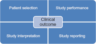
Parameters involved in the Echocardiographic process.
Assessment of Technical Competence
Individual efficiency in echocardiography is not directly related to traditional measures of competence such as training and number of examinations performed. 25 Accordingly, a valid and applicable assessment of technical skills requires an objective and structured tool.26,27 Nielson et al. 25 used a global score and a checklist to evaluate technical skills in TTE. Interestingly, he found that the blinded use of an objective assessment instrument accurately correlates with the levels of performance. More importantly, Norcini 28 reported that assessment of technical and non-technical aspects of medical proficiency require extreme caution given that factors influencing the quality of assessment may be predominant, including reliability, physician relationships, stakes (conflict of interest), and reproducibility.
Classical parameters, such as operator caseload or certifications, have proven to be insufficient to guarantee a high-quality echocardiography.25,29 A structured and objective tool can be implemented in every healthcare institution according to the local needs (ie, audits regarding indications, availability, performance, and diagnostic accuracy). For that purpose, a neutral expert in echocardiography (rater) evaluates randomly chosen series of studies, then blindly re-assesses the same studies to check for intra-rater variability and another expert evaluates blindly the same studies in order to assess the inter-rater variability. 25 Individual competence can be assessed by analysis of multiple parameters, 6 mainly evaluation of a series of studies, 25 together with evaluation of the administrative record (certificates, continuous medical education, experience, evidence of practical ability, testimonial of referring physicians, caseload, etc.).
Table 4 shows a model of objective methodology for individual technical assessment based on “cases, administrative record, series studies.” Minimal evaluation, based on selected cases where selection is made by peers is simply and purely a poor assessment instrument that lacks reliability due to potential conflict of interest. Selected cases could be discussed in medical staff or even in morbidity-mortality meeting in the perspective of quality improvement project (QIP). 28 Technical competence depends also on laboratory infrastructure (logistics, equipment, staffing) and organization (procedures and protocols).
Model of assessment of individual technical competence in Echocardiography.

Quality Improvement Project
Any QIP should aim basically to improve the clinical outcome rather than simply analyzing the echocardiographic product or finding professional errors. In this perspective, it would be unfortunate to have the head of the echo laboratory not sufficiently (or not at all) involved in a continuous QIP. 6 A structured QIP may include a tailored teaching/training program, creation of uniform procedural protocol, and implementation of regular internal audits.6,16 Moreover, a QIP must aim to increase the AUC of echocardiography, keep professional standards, as well as encouraging teamwork for a better clinical benefit.
Johri et al. 18 found that teaching intervention yielded a 40% reduction in inter-observer variability in ejection fraction assessment; similarly, Bhatia et al. 30 applied an educational interventional program that yielded a significant decrease in the rate of inappropriate tests. Besides the human-oriented program, a QIP must aim to improve the technical component of echocardiographic process, ensuring an adequate echographic machine and implementing structured and organized unit.
The objective of any QIP is to monitor, identify, analyze, and treat any dysfunction whether individual or related to the unit. For this purpose, the QIP must use objective and reproducible markers, including the Unit Registry. Registry must be implemented positively for QIP and not only for caseload reporting. Likewise, QIP must aim to develop indices of patient outcome and clinical impact, as well as to develop procedures to determine intra- and inter-observer variability/ discrepancy, to develop standards for interpretation and reporting of echo studies.31–33
Sonographers should seek referral to an advanced level of expertise or even require a complementary paraclinical tests (ie, cardiac magnetic resonance imaging) when it is necessary. In this context, sonographers’ attitude and awareness are of utmost importance in order to keep ethical values and scientific accuracy; otherwise, their behavior may represent endangering skills manifested by reluctance to teamwork with conceited profile. Such profiles are not appropriate to approach clinical challenges, to enhance teamwork and to yield a quality improvement project. The core echocar-diographic training should include a component dealing with this behavioral factor and not only limited to technical product. 34
Conclusion
Appropriate medical practice in echocardiography extends beyond the simple echocardiographic product; it aims to improve the clinical outcome. Patient selection, conformity to AUC, study performance, interpretation, reporting, competence assessment, CUDS, and QIP are essential ingredients to generate a high-quality echocardiographic product with beneficial clinical outcome. In all cases, an echocardiographic study should be comprehensive, accurate, reproducible and addressed to the clinical context. The attitude and profile of sonographers are valuable to improve quality of care, including availability, scientific accuracy, and technical competence. Assessment of technical competence must be conducted in the perspective of QIP and clinical outcome, for the best of the patient and institution.
Author Contributions
Conceived the concept: AK. Analyzed the data: AK. Wrote the first draft of the manuscript: AK. Made critical revisions: AK, GG. All authors reviewed and approved of the final manuscript.
Disclosures and Ethics
As a requirement of publication the author has provided signed confirmation of compliance with ethical and legal obligations including but not limited to compliance with ICMJE authorship and competing interests guidelines, that the article is neither under consideration for publication nor published elsewhere, of their compliance with legal and ethical guidelines concerning human and animal research participants (if applicable), and that permission has been obtained for reproduction of any copyrighted material. This article was subject to blind, independent, expert peer review. The reviewers reported no competing interests.