
Abstract
Hereditary Haemorrhagic Telangiectasia (HHT), or Osler-Weber-Rendu syndrome is an uncommon autosomal dominant multi-organ condition of vascular dysplasias. We describe a 19 year old Indian female who presented with cerebral abscess secondary to paradoxical emboli from pulmonary arteriovenous malformations (PAVMs) associated with HHT. Cerebral, pulmonary, hepatic and gastrointestinal involvement can be life-threatening and it is important to have lifelong follow-ups on these patients.
Keywords
Introduction
The typical patient with HHT has clinical features of recurrent epistaxis, mucocutaneous telangiectases and gastrointestinal bleeding. 1 Besides involving post capillary venules that result in mucocutaneous telangiectases, HHT is associated with the formation of arteriovenous malfomations (AVMs) in the lungs, liver and brain. HHT has a prevalence rate that is as high as 1 in 10,000 in some parts of the world. 2 To make a definite diagnosis according to Curacao criteria, three out of four clinical features are to be present.3,4 These include spontaneous, recurrent nose bleeds, telangiectases at typical sites, visceral AVMs and a first degree relative with known HHT. It is a disease with variable penetrance that has a wide range of clinical presentations.
Case Report
A 19 year old Indian female with no past medical history of note was admitted for complaint of severe headache associated with fever for 5 days' duration. On physical examination, she was alert and orientated, with a fever of 39
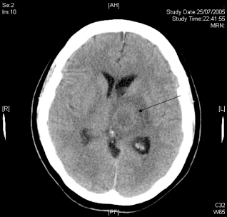
Shows a 2.5 cm vague hyperdense rim-enhancing lesion (arrow) in the left thalamus with minimal mass-effect on 3rd ventricle.

MRI T2 image (2 days later) shows the same rim-enhancing lesion in left thalamus with increased mass effect.
Various investigations were performed to determine the source of the cerebral abscess. Dental review was normal, 2-dimensional trans-thoracic echocardiography did not show any infective endocarditis and otolaryngologist assessment was normal. While recovering in the general ward, the patient had an episode of oxygen desaturation. A CT pulmonary angiogram was done (Fig. 3) to look for pulmonary embolism which was absent. Instead, the scan revealed multiple PAVMs in both lungs. It was postulated that the PAVMs allow right-to-left shunting of paradoxical emboli, resulting in the cerebral abscess.
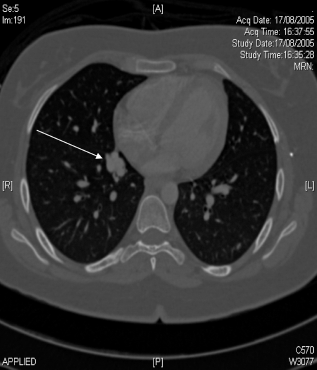
Shows a focal nodular appearance of pulmonary vessels (arrow), representing one of the arteriovenous malfomations in the right lung.
On further questioning, the patient gave a history of recurrent spontaneous epistaxis since the age of ten. On close examination of the patient, a few scattered telangiectases were seen on the tongue and palate. Although there was no positive family history, she fulfilled the criteria for the diagnosis of HHT. 3
Further investigations were performed to exclude AVMs in other sites of the body. MRI of the brain did not reveal any AVMs. CT liver did not reveal any AVMs. Transcatheter embolisation of the PAVMs was performed on 2 separate occasions. Left pulmonary angiogram (Fig. 4) showed 2 lower lobe PAVMs about 1 cm in diameter and multiple embolic coils inserted successfully. Right pulmonary angiogram revealed 4 PAVMs about 1 cm in diameter and embolisation of 3 lesions were done. One PAVM was inaccessible. She was followed up yearly to screen for new development of PAVMs and AVMs from other sites.

Transcather embolisation with a metallic coil (black arrow) into a PAVM (white arrow) located in the right lung.
Discussion
This case illustrates neurological symptoms as the first prominent manifestations of PAVMs and hereditary haemorrhagic telangiectasia. The patient had no family history that would have prompted early screening for characteristic features of HHT and its visceral involvement. Although her epistaxis during her early teens were spontaneous and recurrent, there was no other prominent oral mucosa telangiectases to arouse any suspicion of HHT.
The incidence of cerebral AVMs and PAVMs were estimated to be 10%–15% and 11%–30% respectively; PAVMs are mainly asymptomatic (25%–58%) or present more commonly with respiratory symptoms than embolic phenomenon. 5 The incidence of embolic phenomenon in PAVMs was low, about 0%–25% have cerebral abscess and 11%–55% have transient ischemic attack or stroke. 5 Consequently, neurological symptoms are not common in patients with HHT, they consist of headache, transient ischemic attack, stroke, seizure, intracranial bleed and brain abscess. PAVMs contribute to two thirds of HHT-related neurological symptoms while in the remaining one third, cerebral and spinal AVMs are responsible. 6 Usually, the pulmonary capillaries filter effectively any thrombotic and septic emboli that enter the right side of the heart and prevent their entry into the systemic circulation. The presence of PAVMs allows shunting of these emboli, resulting in embolic manifestation in the brain, as seen in our patient.
HHT is an autosomal dominant disorder. Mutations involving at least two genes were recognized so far: endoglin on chromosome 9 and activin receptorlike kinase 1 on chromosome 12. 5
Large AVMs larger than 1 cm in diameter can cause shunting of blood. These occur commonly in the lung, but the brain, liver and gastrointestinal tract can also be involved. When occurring in the liver, it can result in portal hypertension, high-output heart failure and biliary disease. 7 In the gastrointestinal tract, recurrent bleeding can occur, especially after the age of 30. 5
Our patient had HHT with a severe neurological complication but fortunately, she recovered with no residual neurological deficit. She did not have any cerebral or hepatic AVMs.
It was only in recent time that transcatheter embolisation of PAVMs replaces surgical resection as the treatment of choice. The procedure involves the placement of a metallic coil or balloon to occlude the feeding vessels to PAVMs wth thrombus. It is effective for reducing right-to-left blood shunting, improving hypoxemia and increasing exercise capacity. 8 It may decrease neurological complications. Transcatheter embolisation of PAVMs is considered safe, especially in experienced hands. The success rate has been reported to be over 98% in cumulative series, with no report of mortality related to procedure. 9 Pleurisy, paradoxical embolism and balloon deflation are some possible complications. Currently, transcatheter embolisation is recommended for PAVMs with feeding arteries greater than 3 mm in diameter. 10 Surgical resection is indicated for patients who had a persistent right-to-left shunt following embolisation of all significant PAVMs. Lung transplant is also considered feasible in patients with diffuse disease.
Pulmonary angiography is considered the gold standard in detection of PAVMs. But three other non-invasive methods are currently available to detect PAVMs:
Radiography–-chest radiograph or CT thorax
Detection of hypoxemia by pulse oximetry or arterial blood gas
Detection of right to left shunt–-100% inspired oxygen breathing method or contrast echocardiography or radionuclide scanning
In a group of 105 patients with HHT, Cottin and colleagues 11 did a comparison of the accuracy of a group of non-invasive methods to detect PAVMs against CT thorax and/or pulmonary angiography. They recommended a screening algorithm that uses chest radiograph and contrast echocardiography, followed by CT thorax if either test is positive. Screening directly by CT thorax is also an effective alternative.
Our patient had been followed-up over the last few years with regular pulse oximetry monitoring and serial CT thorax scans. These had been normal so far and the follow-up is intended to be lifelong. Repeated screening tests for brain and liver AVMs are not recommended as there is no evidence that they increase in size over time.
Besides screening for AVMS in the various organs, management of HHT also include treating the complications that may arise during the patient's lifetime. Telangiectases, if comestically undesirable, can be treated with topical agents or laser ablation. Significant or recurrent epistaxis can be treated with cauterization, laser ablation, septal dermatoplasty, course of estrogen or transcather embolisation of arteries feeding nasal mucosa. Bleeding in the gastrointestinal tract can be treated by endoscopic heater probes or laser. Patient education is essential. The patient was told of the need for antibiotic prophylaxis prior to any dental or surgical procedure. Besides screening the rest of her family members for this condition, we also provided genetic counseling to the patient as she is of child-bearing age.
Conclusion
This is an interesting case of brain abscess of which further investigations led to a diagnosis of a rare systemic condition of HHT and subsequent prevention of further life threatening complications. It serves to remind us the need to be thorough in our history taking and physical examination when seeing a patient. In addition, the case also emphasizes the importance of lifelong follow-ups of systemic conditions that may have unpredictable clinical outcome and complications.
Footnotes
This manuscript has been read and approved by all authors. This paper is unique and is not under consideration by any other publication and has not been published elsewhere. The authors and peer reviewers of this paper report no conflicts of interest. The authors confirm that they have permission to reproduce any copyrighted material.