
Abstract
The incidence of ectopic pregnancy is approximately 1.3∼2% of all pregnancies, and more than 90% of ectopic pregnancies are detected in the ampulla of the fallopian tube. Ectopic pregnancy occurring in tubal stump after tubectomy is extremely rare, and the frequency of tubal stump pregnancy is approximately 0.4% of all pregnancies. We report one of these rare cases of ectopic pregnancy in a 26-year-old Japanese woman, gravida 4, parity 1. She had undergone laparoscopic tubectomy because of a tubal pregnancy two years ago. She was presented to our hospital with a positive pregnancy test, but no gestational sac was detected in the uterus by echography, even though the level of human chorionic gonadotropin (hCG) in the blood was elevated to 8,900 mIU/mL. Laparoscopic surgery for ectopic pregnancy was performed. During surgery, the position of the pregnancy was found to be in the tubal stump, where tubectomy had already been performed, and the gestational sac was successfully removed. After the surgery, the condition of the patient uneventfully improved and she was discharged from the hospital three days after the surgery. The diagnosis of tubal stump pregnancy is more difficult than that of the more common positions of an ectopic pregnancy in the fallopian tube, and so it is more important to carefully examine the patients with suspected ectopic pregnancy. Laparoscopic surgery is one of the options for tubal stump pregnancy if diagnosed early and if the condition of the patient is stable.
Introduction
Ectopic pregnancy is a major health problem for women in many countries. The prevalence of ectopic pregnancy is high in societies in which women delay childbearing, with increased use of assisted reproductive technology (ART) and with increased incidence of pelvic inflammatory disease caused by chlamydia and gonococcal bacteria. The unique anatomic location of the tubal stump pregnancy sometimes results in delayed diagnosis. The risk of rupture of the uterus tends to increase beyond 12 weeks of amenorrhea, and the rupture of the ectopic part occurs in 20% of ectopic pregnancies beyond 12 weeks of gestational age. Developing better methods for earlier diagnosis would decrease morbidity and increase the chance of successful laparoscopic surgery.
In this paper, we present the case of a patient with tubal stump pregnancy. The patient underwent successful laparoscopic surgery, and experienced an uneventful recovery.
Case Reports
The patient was a 26-year-old Japanese woman, gravida 4, parity 1, who had a medical history of laparoscopic surgery for right tubal pregnancy two years ago, and no remarkable family history.
She was presented to our hospital with a positive pregnancy test. No gestational sac was detected in the uterus by transvaginal sonography, even at 6 weeks 0 days' gestational age. However, the level of human chorionic gonadotropin (hCG) in the patient's blood was elevated to 8,900 mIU/mL, and a gestational sac-like mass was found outside of the uterus by echography (Fig. 1). Based on these test results, ectopic pregnancy was strongly suspected, and the patient was suggested to undergo laparoscopic surgery.

The lesion of tubal stump pregnancy appears in the fallopian tube at the left side of uterus.
The operative findings revealed a mass in the right tubal stump (Fig. 2), and we diagnosed it as tubal stump pregnancy. Before resection of the ectopic part, we injected vasopressin surrounding the ectopic lesion to decrease blood flow. The ectopic part was removed with an advanced bipolar sealing device, LigaSure® (Covidien, Manhattan), and the operative wound was sutured with absorbable VICRYL® (Ethicon, New Jersey) for uterine wall repair (Fig. 3). We were not able to find any chorionic villi macroscopically (Fig. 4), but pathological findings revealed chorionic villi around the removed tubal stump, confirming the diagnosis of tubal stump pregnancy (Fig. 5).

The laparoscopic finding of the ectopic part at the right tubal stump of the uterus.

The wound after resection of the ectopic part is sutured with VICRYL, with only a small amount of bleeding.
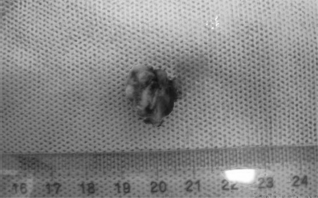
Nonvisibility of the chorionic villi macroscopically in the removed lesion.

The chorionic villi detected microscopically in the removed ectopic part. (
After the surgery, the level of hCG in the blood decreased and the patient recovered uneventfully. The patient was discharged three days after the operation.
Discussion
Ectopic pregnancy is a major threat to the life of women of reproductive age. Recently, the frequency of ectopic pregnancy has tended to increase because of delayed childbearing, the increased use of ART, and an increase in sexually transmitted diseases. 1 It is reported that the incidence of ectopic pregnancy is approximately 1.3%–2% of all pregnancies. 2 More than 90% of ectopic pregnancies are detected in the tubal ampulla, and approximately 2.5% of cases occur in the interstitial position. 3 Ko et al reported that tubal stump pregnancy after tubectomy is extremely rare, with a prevalence of about 0.4%. 1
There are two hypotheses for the pathogenesis of tubal stump pregnancy. One is the external chemotactic theory, which postulates that a foramen is formed at the tubal stump and a fertilized ovum coming from the intra-abdomen implants into the tubal stump portion.1,4 The other theory is that an embryo coming from the normal contralateral fallopian tube implants in the tubal stump.5,6 In this case, we could not see any foramen on the tubal stump, and hence the latter theory is applicable.
Ultrasonographic examination is effective for the diagnosis of tubal stump pregnancy. However, in some cases, the diagnosis of tubal stump pregnancy is difficult because the tubal stump portion is near the ovary, and hence, the ovarian follicle is mistaken for a tubal stump pregnancy. Moreover, it is thought that many doctors pay less attention to the tube in which patients have already undergone tubectomy because of ectopic pregnancy. Lau and Tulandi proposed three sonographic criteria for interstitial and tubal stump pregnancies: (1) an empty uterine cavity, (2) a chorionic sac seen separately and
The main treatment for tubal stump pregnancy is surgery and conservative management using methotrexate. Lau and Tulandi reported that the overall success rates in surgical treatment reached 100% and that of methotrexate management was 83%. 7
The operation method for tubal stump pregnancy is almost the same as that of interstitial pregnancy. The difficulty level of laparoscopic operation for interstitial and tubal stump pregnancy is higher than that of common laparoscopic tubectomy, and hence, the selection of operative method depends on the surgeon's preference and expertise. There are several reports of successful laparoscopic operation for interstitial and tubal stump pregnancy using an advanced bipolar device and injecting diluted vasopressin into the uterus.9–11 Sherer et al recommend clamping the adjacent uterine wall to the interstitial pregnancy with long-jaw forceps before incising the cornua. 12 On the other hand, there are reports of using hysteroscopic surgery for interstitial and tubal stump ectopic pregnancy. 13 However, the efficiency and long-term prognosis for selecting hysteroscopic surgery are unknown. In this case, we injected vasopressin into the uterus surrounding the ectopic part and removed the ectopic lesion from the tubal stump using an advanced bipolar sealing device, LigaSure®. We successfully performed the operation for this patient with tubal stump pregnancy.
Any subsequent pregnancy after operation for tubal stump pregnancy should be followed up carefully and cesarean delivery at term may be safer and help decrease the risks of uterine rupture during labor.
In summary, we successfully treated a case of tubal stump pregnancy with laparoscopic surgery. Laparoscopic surgery might be the first-line treatment for a hemodynamically stable patient with interstitial pregnancy of a small size. Sometimes, the accurate diagnosis for this type of ectopic pregnancy is difficult; therefore, we have to pay much attention to the possibility of tubal stump pregnancy when we diagnose the ectopic pregnancy.
Author Contributions
Conceived and designed the experiments: MN, YM, YK, KT, HN. Analyzed the data: MN, YM, YK, KT, HN. Wrote the first draft of the manuscript: MN, YM, YK, KT, HN. Contributed to the writing of the manuscript: MN, YM, YK, KT, HN. Agree with manuscript results and conclusions: MN, YM, YK, KT, HN. Jointly developed the structure and arguments for the paper: MN, YM, YK, KT, HN. Made critical revisions and approved final version: MN, YM, YK, KT, HN. All authors reviewed and approved of the final manuscript.