
Abstract
To evaluate collagenase inhibitors for the treatment of osteoarthritis and to correlate them with clinical pathology, canine cartilage explant and anterior cruciate ligament transection (ACLT) models were examined by quantifying the CII neoepitope (CIINE). This peptide is a putative marker for collagenase-specific type II collagen (CII) degradation, which is a critical step in osteoarthritis pathology.
The concentration of CIINE in supernatants of canine cartilage explants showed increase upon IL-1β—stimulation and collagenase inhibitors suppressed this elevation of CIINE. In the canine ACLT model, levels of CIINE in urine (uCIINE) increased as lesions of knee joint cartilage developed and decreased in response to collagenase inhibitors.
Our results suggest that CIINE reflects collagenase-specific CII degradation in canine explants and whole bodies. It is anticipated that these data will establish a tool for clarifying and bridging the efficacy and mechanism of collagenase inhibitors at the preclinical stage of drug discovery.
Introduction
Osteoarthritis (OA) is the most common type of joint disease and is characterized by progressive degeneration and erosion of articular cartilage. 1 It is well known that collagenases, a subset of matrix metaloproteinases (MMPs) produced by chondrocytes and synovial cells,2–7 are predominantly responsible for the cleavage of type II collagen (CII) in cartilage extracellular matrix (ECM).1,8–16,17 The human collagenases MMP-1 and MMP-13 are critical to the initiation of type II collagen breakdown because they perform the rate-limiting steps in CII degradation during OA and represent an important target of disease-modifying OA drugs (DMOADs). However, to date, poor accuracy and target specificity of assessments of cartilage degradation and discrepancies of collagenase profiles between humans and rodent models have prevented the development of efficacious DMOADs.18–21
The use of biomarkers of cartilage destruction throughout the stages of drug discovery has been advocated 20 as a solution to these issues. CII neoepitope (CIINE) has been said to be a promising putative collagenase-specific biomarker of CII degradation.8–10,17 This epitope (GPPGPQG) is located in the N-terminal vicinity of a collagenase-specific cleavage site on the CII molecule, and most peptides with this epitope are excreted in urine.9,11 Importantly, elevated urine CIINE (uCIINE) levels have been observed in OA patients.22,23
To establish biomarker usage in animal models with collagenase profiles that are consistent with humans, we validated CIINE as a marker of cartilage degradation in canines. Therefore, the advantages of using biomarkers and the potential of CIINE to surrogate cartilage breakdown will be discussed for every stage of drug discovery.
Methods
Animals
All animals in this study were obtained from Japan SLC Inc. (Hamamatsu, Japan) and were maintained in accordance with the rules and regulations for the care and use of experimental animals that are stipulated by the Shionogi Animal Use and Care Committee. IRB approval for this research was obtained.
Reagents
Unless otherwise stated, all the reagents were purchased from Life Technologies Corporation (Carlsbad, CA, USA). S-3536 and marimastat were prepared at Pharmaceutical Research Laboratory, Shionogi & Co., Osaka, Japan.
Anterior cruciate ligament transection and urine sampling
It was confirmed that the background levels of uCIINE were low and stable in animals > 35 months old. Three 68-month-old dogs received anterior cruciate ligament transection (ACLT) on the right knee joint under general anesthesia as described in previous studies.24–26 In brief, the anterior cruciate ligament was transected from the right hind limb, ensuring that no other structure was damaged. The procedure was confirmed to be successful using a positive anterior drawer sign. Fresh urine from each dog was collected every 2 weeks. The supernatants of urine samples were collected after centrifugation and stored at −80 °C until uCIINE measurements were carried out. Six animals were orally administered 30 mg/kg S-3536 (twice a day) between 70 and 74 days after the surgery and were sacrificed on day 90 for histological observations of knee joint injury. Articular cartilage was stained with india ink for macroscopic observations of cartilage degradation. 27 To estimate histological changes before day 90, 3 other ACLT-treated dogs were sacrificed on day 11.
Canine explant
The medium used for explant experiments were prepared as follows. Cleaning medium 1 comprised DMEM, Dulbecco's modified Eagle's median comprised 100 units/mL penicillin, 100 μg/mL streptomycin, 2.5 μg/mL amphotericin B, and 50 μg/mL gentamycin. Cleaning medium 2 comprised DMEM containing 1000 units/mL penicillin and 1000 μg/mL streptomycin. Culture medium comprised AIM-V supplemented with 100 units/ml penicillin, 100 μg/ml streptomycin, 1% l-glutamine, 1% ITS (Sigma-Aldrich, St. Louis, USA), 0.1 mg/mL BSA (Sigma) and 50 μg/mL l-ascorbic acid (Wako Pure chemical Industries, Osaka, Japan).
Adult canine (15–36 months old) articular cartilage was obtained from the femur chondyler and tibia bones of knee joints shortly after sacrifice under anesthesia. Cartilage was soaked in cleaning medium 1 for 30 minutes followed by cleaning medium 2 for 30 minutes, and then minced into pieces of 1 to 2 mm in diameter. Approximately 40 mg of minced samples were precultured on a 48-well cell culture plate with culture medium at 37 °C and 5% CO2 for 2 days before conducting cartilage stimulation experiments.
Stimulation of cartilage explants was conducted by replacing culture supernatant with fresh media containing 50 ng/mL human IL-1β (R&D Systems, Minneapolis, USA). Spent medium was collected every 3 or 4 days and stored at −80 °C until analysis. In MMP inhibitor experiments, culture medium was supplemented with the inhibitors S-3536 or marimastat.
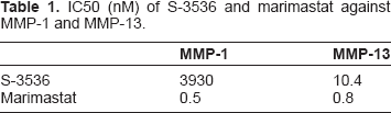
Inhibition of MMP-1 and MMP-13 was measured as described below (Table 1). Human MMP-1 was purchased from Yagai Co: (Yamagata, Japan). The MMP13 catalytic domain (CD) construct encompassing Tyr 105 to Glycin 206 of human MMP-13 was generated in a bacterial expression system. 28 The substrate MOCAc-Pro-Leu-Gly-Leu-A2pr(Dnp)-Ala-Arg-NH2 (3163-v) was purchased from Peptide Institute (Osaka, Japan). Enzymatic activity was analyzed using a previously reported method. 29 The half inhibitory concentration (IC50) of each compound was calculated as that required to inhibit enzymatic activity to 50%.
IC50 (nM) of S-3536 and marimastat against MMP-1 and MMP-13.
Measurement of CIINE and active MMP-13
To measure CII-derived peptide CIINE (Col2-3/4Cshort) concentration by sandwich enzyme-linked immunosorbent assay (ELISA), a mouse monoclonal antibody for intrachain CII 6G4 and the mouse monoclonal antibody 20A10, which recognizes the CIINE generated by collagenase, were used as detector and capture antibodies, respectively 30 The concentration of CIINE in urine samples was normalized to creatinine concentrations. Quantification of urine creatinine was performed using a Wako creatinine test (Wako Pure chemical Industries, Osaka, Japan).
To detect activated MMP-13 by sandwich ELISA, the mouse monoclonal antibody 14D10, which is specific to the activated MMP-13 catalytic domain, and anti-MMP-13 mouse monoclonal antibodies (Daiichi-Fine Chemical, Toÿama, Japan, Clone 181–15A12) were used as detector and capture antibodies, respectively.
Statistical analysis
The mean and standard deviation (SD) of each variable were calculated. Data was analyzed using Student t test or analysis of variance (ANOVA) with Graph-Pad Prism. P values less than 0.05 were considered significant.
Results
Time-dependent changes in CIINE and MMP-13 levels
In IL-1β—stimulated explants, increase in CIINE levels was time-dependent, with a 24-fold increase at 0 to 4 days and 12 to 14 days of culture. In unstimulated samples, CIINE levels increased only 8-fold compared with the initial level (Fig. 1A). In addition to CIINE, active MMP-13 was induced by IL-1β in a time-dependent manner, and in unstimulated samples it increased marginally in clear increments (Fig. 1B).

IL-1β caused time-dependent increases in CIINE and active MMP-13 levels in canine cartilage explants.
After 14 days of culture, CIINE levels in unstimulated samples increased sharply (data not shown) but were maintained in IL-1β—stimulated samples. To exclude this IL-1β—independent event, experiments using MMP inhibitors were conducted before 12 days of culture.
Effects of MMP inhibitors on IL-1β—induced CIINE production
We tested 2 different types of MMP inhibitors, marimastat, and S-3536. Marimastat is a powerful broad-spectrum inhibitor of MMPs such as MMP-1, −2, −3, −8, −9, −12, −13 and −14. Marimastat demonstrated strong inhibition of MMP-1 and MMP-13 activity, with IC50s of 0.46 nM and 0.8 nM, respectively (Table 1). In contrast, S-3536 was selective for MMP-13, with IC50 s of 3930 nM and 10 nM against MMP-1 and MMP-13, respectively.
In subsequent experiments, inhibition of IL-1β—induced cartilage degradation was examined after adding S-3536 or marimastat to the media throughout the cultural period (Fig. 2). Data from each of the supernatants collected on days 4 to 8, 8 to 12, and 12 to 14 revealed similar trends. In these experiments, marimastat inhibited CIINE production to below control levels, and S-3536 at >50 μM almost abrogated IL-1β—induced CIINE production but had no significant effect at 10 μM.

S-3536 and marimastat inhibited IL-1β—induced CIINE production.
In pharmacological tests, S-3536 at 100
Macroscopic evaluation and biochemical detection of cartilage destruction caused by ACLT
Eleven days after ACLT surgery in dogs, the surfaces of articular cartilage were slightly stained with india ink, indicating damage to the cartilage matrix. Three months after the surgery, this disrupted india ink— stained area was expanded and outstanding (Fig. 3).

India ink staining visualized ACLT induced injury in canine articular cartilages.
Consistent with the course of cartilage degeneration, uCIINE levels increased in all dogs that received ACLT, whereas in sham-treated animals, uCIINE remained at basal levels (Fig. 4). Urine CIINE levels were elevated 42 days after ACLT and continued to increase throughout the observed period until day 81. When S-3536 was administered on day 70, uCIINE levels decreased suddenly but restored to baseline levels in 4 days.
Discussion
Treatment with collagenase inhibitors is often stopped due to insufficient effect or side effects such as musculoskeletal syndrome (MSS).21,31–33 Considering this, we focused on the insufficiencies of nonclinical drug evaluations, particularly in animal experiments. Cartilage damage is commonly evaluated by macro observations with india ink. This method is only poorly quantitative of the intensity of damage and provides no information about target specificity. Because animals must be sacrificed to assess cartilage damage, it is impossible to monitor pathogenesis in individuals over time and thus to optimize the timing of treatments.

The uCIINE levels in ACLT-treated dogs showed longitudinal increase.
To address this issue, biomarkers for collagenase-dependent cartilage damage have been proposed and several biochemical and radiographical candidates have been investigated. 20 In this study, we investigated CIINE because it reflects collagenase-specific CII breakdown and is quantitatively and noninvasively measurable in urine.8–10,17 With these advantages, CIINE may provide a tool for determining cartilage damage that limits side effects and allows timely quantitative assessments of collagenase inhibitors in vivo.
Another issue regarding the evaluation of collagenase inhibitors is the difference in collagenase profiles between humans and widely used rodent models of OA. Critically, while MMP-13 is expressed in both humans and mice, rodents lack MMP-1 and are thus inappropriate for specific demonstration of collagenase inhibitors intended for human use. On the other hand, canines express both MMP-1 and MMP-13, and the well-established canine ACLT model bears morphologic, metabolic, biochemical, and biomechanical features and osteophyte formation in the articular cartilage of the unstable knee that mimics those of human OA. 24,26,34–36 Moreover, damaged cartilage and synovial joint fluid are CIINE-positive in ACLT dogs. 25
To validate CIINE as a cartilage degradation marker in canines, we conducted CIINE and MMP-13 ELISA 30 in cartilage explants. Similar to previous studies using immortalized or primary human and bovine chondrocytes,37–40 CIINE and MMP-13 were increased in response to IL-1β. Although the levels of MMP-1 were not measured in this study, MMP-1 is also believed to contribute to IL-1β—induced CII degradation, which has been proved in previous studies.37,41
In this study, CII degradation in canine cartilage explants was inhibited by the MMP inhibitors marimastat and S-3536. Marimastat is a well-known multiple MMP inhibitor and is highly effective against both MMP-1 and MMP-13. On the other hand, S-3536 was designed to selectively inhibit MMP-13, though it is not completely specific (Table 1). In canine explants, both inhibitors suppressed the IL-1β—induced increase in CIINE, but the effects of marimastat were more clearly dose-dependent than those of S-3536. S-3536 almost completely inhibited CIINE production at 100 μM, whereas marimastat completely inhibited CIINE production at only 10 μM. Assuming similar pharmacokinetic properties in explant cultures, these data may reflect differing collagenase specificities of marimastat and S-3536. Although marimastat was more potent as an inhibitor of MMP-1 and MMP-13 than S-3536, this difference was more marked with respect to MMP-1 alone (Table 1). Therefore, we speculate that superior inhibition of IL-1β—induced CII destruction by marimastat primarily reflects inhibition of MMP-1. While the degree to which IL-1β controls cartilage degradation remains unknown, its involvement is indicated in numerous studies.37,41–43 Hence, while MMP-13 cleaves CII more efficiently than MMP-144,45 and is highly expressed in OA chondrocytes, 19 it appears that MMP-1 is also important in CII degradation.
We have successfully demonstrated the use of CIINE in monitoring degradation of CII and evaluating MMP inhibitors in canine explants, and we have confirmed its application to cartilage damage in ACLT-treated dogs. In contrast to cartilage explants, the whole body comprises numerous tissues abundant in CII. Hence, lesion-specific variations in uCIINE levels may be obscured by considerable physiological background. To reduce this background, we experimented in adult dogs with low and stable uCIINE, and observed markedly increased uCIINE after ACLT (Fig. 4). These data provide significant evidence of CIINE release and urinary excretion from damaged cartilage tissue. In addition, noninvasive measurements of uCIINE can quantify the onset and progression of collagenase-specific cartilage degradation such that the timing of inhibitor treatment can be optimized. We expect this simple noninvasive diagnostic tool to succeed assessments of macroscopic scores.
Conclusion
We believe that our study clearly demonstrates the feasibility of drug efficacy investigations that are based on the presence of CIINE in canine explants and ACLT models. Using dogs as an animal model is beneficial to correlate the efficacy to the clinical use because they possess the same collagenase as humans, MMP-1 and MMP-13, unlike rodents, which have only MMP-13 as collagenase. This development will substantially contribute to the refinement of drug evaluations and will establish proof of mechanism and efficacy of collagenase inhibitors at the preclinical stage. With further studies on the validity of uCIINE as a clinical surrogate marker in clinical OA and canine ACLT, preclinical studies using CIINE may predict clinical effectiveness of drug candidates for OA.
Author Contributions
Conceived and designed the experiments: SM, MT, AN, MK, AY, JO, T. Tsuji, T. Takahashi, and TT-M. Analysed the data: SM and MT. Wrote the first draft of the manuscript: SM. Contributed to the writing of the manuscript: SM and TT-M. Agree with manuscript results and conclusions: SM, MT, TT-M, YM, and NF. Jointly developed the structure and arguments for the paper: SM, MT, TT-M, YM. Made critical revisions and approved final version: SM, MT, TT-M, and NF. All authors reviewed and approved of the final manuscript.
Funding
Author(s) disclose no funding sources.
Competing Interests
Author(s) disclose no potential conflicts of interest.
Disclosures and Ethics
As a requirement of publication the authors have provided signed confirmation of their compliance with ethical and legal obligations including but not limited to compliance with ICMJE authorship and competing interests guidelines, that the article is neither under consideration for publication nor published elsewhere, of their compliance with legal and ethical guidelines concerning human and animal research participants (if applicable), and that permission has been obtained for reproduction of any copyrighted material. This article was subject to blind, independent, expert peer review. The reviewers reported no competing interests.
Footnotes
Acknowledgments
The authors would like to thank Enago for the English language review.