
Abstract
Common genetic variants mapping to two distinct regions of RADS1B, a paralog of RADS1, have been associated with breast cancer risk in genome-wide association studies (GWAS). RADS1B is a plausible candidate gene because of its established role in the homologous recombination (HR) process. How germline genetic variation in RADS1B confers susceptibility to breast cancer is not well understood. Here, we investigate the molecular function of RADS1B in breast cancer cell lines by knocking down RADS1B expression by small interfering RNA and treating cells with DNA-damaging agents, namely cisplatin, hydroxyurea, or methyl-methanesulfonate. Our results show that RAD51B-depleted breast cancer cells have increased sensitivity to DNA damage, reduced efficiency of HR, and altered cell cycle checkpoint responses. The influence of RAD51B on the cell cycle checkpoint is independent of its role in HR and further studies are required to determine whether these functions can explain the RADS1B breast cancer susceptibility alleles.
Introduction
In mammalian cells, the repair of DNA double-strand breaks (DSBs) can occur via two distinct mechanisms: non-homologous end joining (NHEJ) and homologous recombination (HR).1,2 HR uses a sister chromatid or homologous chromosome as a template during repair and thus is considered an error-free mechanism for repairing DSBs that arise spontaneously or in response to DNA-damaging agents. 3 Previously, it has been shown that RAD51 protein forms a nucleofilament with single strand DNA as part of the DNA paring and strand exchange steps of HR.4,5
There are five RAD51 paralogs in humans that share 20–30% DNA sequence identity and paralogous protein sequences have been identified in vertebrates: RAD51B,6–8 RAD51C, 9 RAD51D,8,10,11 XRCC2,12–14 and XRCC3.12,15 Two distinct protein complexes, RAD51B-RAD51C-RAD51D-XRCC2 (BCDX2) and RAD51C-XRCC3 (CX3), 16 have been shown to form ring-shaped structures that bind to Y-shaped replication-like intermediates and synthetic Holliday junctions by electron microscopy 17 during DNA replication and resolution of HR intermediary structures. 18
In 1994, mutations in the BRCA1 gene were linked to breast cancer susceptibility in high-risk families. 19 Subsequent work has shown that BRCA1 mutations contribute to breast cancer risk by disrupting distinct DNA damage response mechanisms including HR, cell cycle checkpoint arrest, and NHEJ.20,21 Targeted sequencing studies of the RAD51-paralogs have established moderately penetrant germline mutations associated with either breast or ovarian cancer risk (RAD51B, RAD51C, RAD51D, XRCC2, and XRCC3).22–26
For common genetic variants with small effect sizes, GWAS of breast cancer have conclusively identified common single nucleotide polymorphism markers that map to an intronic region of the RAD51B gene. 27 Further investigation of this region has shown an association with the triple-negative subtype in women of European28,29 and African American 30 ancestry, as well as mammographic density measures which predict breast cancer risk. 31 A subsequent breast cancer GWAS has revealed a second, independent signal, marked by a common variant, 335 Kbp away from the primary signal in the 5’ end of the RAD51B gene. Notably, this new region is also associated with male breast cancer. 32
RAD51B is expressed across a spectrum of human tissue. Moreover, the change in measured transcripts upon exposure to DNA-damaging agents has been studied in mammalian cell lines, with only a small effort devoted to breast cancer cell lines. For instance, ionizing radiation in both human foreskin fibroblasts 7 and MCF7 cells, a breast cancer cell line, 33 resulted in an increase in the level of RAD51B transcripts. Studies of ultraviolet (UV) irradiation of CHO cells revealed consensus binding sites for both AP2 and p53 proteins that may regulate RAD51B in response to radiation. 34 RAD51B–/– cells generated in the hyper-recombinogenic chicken DT40 cell line display elevated DNA-damage sensitivity and chromosomal aberrations, whereas there is a reduction in sister chromatid exchange, gene targeting, and DNA damage-dependent RAD51 protein foci formation. 35 Studies using HCT116 colorectal cancer cell lines showed similar results. 36
It is also notable that RAD51B has been shown to influence cell cycle progression. Overexpression of the wild-type RAD51B protein in the p53 mutant CHO cell background induced G1 delays, 37 while knockdown of the gene in HeLa cells by small interfering RNAs (siRNAs) delayed cell cycle progression. 38 In addition, RAD51B has been implicated in the cellular response to platinum drug treatment via the cell cycle checkpoint response as opposed to the DNA repair processes; the increase in RAD51B protein foci (formed in response to DNA damage and thought to represent nuclear domains for HR repair) has been postulated to mediate cell cycle arrest in response to the platinum drugs, oxaliplatin and cisplatin. 39
We have conducted a series of studies in breast cancer cell lines focused on the RAD51B response to DNA damage, specifically examining cell cycle regulation and HR efficiency. We evaluated three breast cancer cell lines, representing different subtypes of breast cancer based on expression of estrogen receptor (ER), progesterone receptor (PR), and HER2. Specifically, BT549 is a triple-negative breast cancer cell line, and MCF7 and T47D are ER-positive, PR-positive, and HER2-negative. We also note that they are polymorphic at the rs999737 risk allele associated with breast cancer; both BT549 and MCF7 are homozygous for the risk allele, and T47D is heterozygous for the risk allele.
Materials and Methods
Cell culture and transfection conditions
Human breast carcinoma cell lines were grown in DMEM (MCF7 and MCF7-DRGRP) or RPMI (BT549 and T47D) supplemented with 10% fetal bovine serum (Life Technologies), 20 mM HEPES, 100 μg/ml streptomycin, and 100 units/ml penicillin (Sigma). The following siRNAs were used for knockdown experiments: AllStars Negative Control siRNA (SI03650318, Qiagen) and siGENOME Human RAD51B siRNA SMARTpool (M-011373, Thermo Scientific). Transfection of siRNA was carried out sequentially using Lipofectamine 2000 (Life Technologies) following the manufacturer's recommendations. Briefly, 100 nM of control or RAD51B siRNA was transfected upon seeding 2 × 10⁶ cells in a 35-mm dish, and then repeated after 24 hours. RNA extractions were carried out using the RNeasy Mini Kit (Qiagen) 48 hours after the initial transfection. cDNA was synthesized using SuperScript® III Reverse Transcriptase Kit (Life Technologies) according to the manufacturer's protocol using 5 ug of total RNA and random hexamers.
Western blots
Protein extracts for Western blots were prepared with RIPA buffer (Santa Cruz Biotechnology, Inc.) containing complete protease inhibitor cocktail (Roche). Protein concentration was measured using the NanoOrange Protein Quantification Kit (Life Technologies). Thirty micrograms of protein were loaded onto NuPAGE 4–12% Bis-Tris protein gels (Life Technologies), electrophoresed, and transferred onto nitrocellulose membranes using the iBlot Gel Transfer Device (Life Technologies). The membrane was then blocked with 5% milk in TBST for 1 hour at room temperature. The following antibodies were used for immunodetection: anti-Rad51B (Abcam: ab124675, 1:500), anti-ActB (Protein-Tech Group: 20536-1-AP), and HRP-conjugated goat anti-rabbit IgG (Proteintech Group: SA00001-2, 1:2000).
Measurement of HR frequency by DR-GFP assay
MCF7DR-GFP cells, and pCBASce, were provided by Dr. Maria Jasin. 2 MCF7DR-GFP cells were transfected with siRAD51B, then co-transfected with 1 ug of siRAD51B and 2 ug of pCBASce or pCBA plasmid 24 hours later using Lipofectamine 2000 (Life Technologies). Cells were harvested after 72 hours and GFP measurements were carried out using a flow cytometer (FACSCalibur; BD Biosciences).
Cell viability assay
Twenty-four hours after transfection with siCON or siRAD51B, 5–10 × 103 cells were seeded in 96-well plates and incubated with 0, 40, or 100 uM cisplatin (CDDP) (Sigma-Aldrich) or 0, 0.4, or 1 mM hydroxyurea (HU) (Sigma-Aldrich). Cell proliferation assays were conducted after 3 days of continuous drug treatment using WST-1 (Roche) according to manufacturer's instructions.
Colony formation assay
MCF7 and T47D cells were transfected with siCON or siRAD51B. After 24 hours, cells were treated for 24 hours with no drug (ND), 100 uM CDDP, or 2 mM (HU). After treatment, cells were washed three times with PBS, trypsanized, and suspended in the appropriate growth medium and 1000 cells were plated. After 14 days, colonies were washed with PBS, fixed with ice cold methanol for 10 minutes, and then stained with 0.5% crystal violet solution in 25% methanol. Only colonies containing more than 50 cells were considered. The fraction of surviving colonies for each drug treatment was calculated by normalizing the number of colonies with siCON and ND treatment.
Real-time polymerase chain reaction
UBC, YWHAZ, and GAPDH were used to normalize RAD51B expression in real-time polymerase chain reaction (RT-PCR) experiments. For UBC (forward: ATTTGGGTCGCGGTTCTTG, reverse: TGCCTTGACATTCTCGATGGT); for YWHAZ (forward: ACTTTTGGTACATTGTGGCTTCAA, reverse: CCGCCAGGACAAAACAGTAT); and for GAPDH (forward: TGATGACATCAAGAAGGTGGTGAAG, reverse: TCCTTGGAGGCCATGTGGGCCAT). Power SYBR Master Mix (Life Technologies) and two sets of primers were used to measure RAD51B expression: ex4.1F (GCACAAAGGTCTGCTGATTTC), ex5.1R (CCCATGTTGGTGGGTAATGT) and ex2.1-15 (TGGGTAGCAAGAAACTAAAACGA), ex3-26 (GGCCCTGCTGACCATACATA).
Microarray and PCR array analysis
The 3′ IVT Express Kit (Affymetrix) was used to amplify and label RNA samples (T47D transfected with siCON or siRAD51B treated with HU) which were subsequently hybridized to the GeneChip Human Genome U133 Plus 2.0 Array (Affymetrix). Background was subtracted, and quantile-normalized data were analyzed using GenomeStudio (Illumina) and Gene Set Enrichment Analysis (GSEA; Broad Institute) software packages. Identical samples were analyzed using the Human DNA Damage Signaling Pathway RT2 Profiler PCR Array (Qiagen) according to the manufacturer's instructions. Data are available at GEO, accession GSE56940.
Cell cycle analysis
CDDP- or HU-treated cells were trypsinized at indicated time points, washed in PBS, and then fixed and permeablized in ice-cold 70% ethanol. Cells were stained with propidium iodide (50 ug/ml) and treated with 100 ug/ml RNaseA for 30 minutes at 30°C, and then analyzed by flow cytometry (FACSCalibur; BD Biosciences). Cell cycle data were analyzed by the Watson (pragmatic) cell cycle model, which does not make any assumption about the S-phase distribution (FlowJo 7.6.5 software, Treestar).
Data analysis
The differences between groups were determined by two tail unpaired t-tests using Excel. Data were considered significant for p < 0.05 (marked as single star). Flow cytometry data were analyzed with FlowJo 7.6.5 analysis software from Tree Star.
Results
Depletion of RAD51B increases sensitivity to DNA-damaging agents
The efficiency of RAD51B knockdown by siRNA was evaluated in three breast cancer cell lines, BT549, MCF7, and T47D. Relative to control siRNA, we observed a 61.4%, 92.7%, and 74.9% reduction of RAD51B transcript for MCF7, T47D, and BT549 cell lines, respectively, 48 hours post transfection. Western blot analysis confirmed the decrease in protein expression (Fig. 1A). The effect of the RAD51B knockdown on DNA damage response was evaluated following treatment with CDDP or HU, using a standard colony formation assay, which measures anchorage-independent cell growth. 40 HU induces stalled DNA replication forks, which are mainly repaired by the HR pathway. CDDP induces intra- and inter-strand DNA crosslinks that are repaired primarily by the nucleotide excision repair (NER) pathway. 41 MCF7, T47D, and BT549 were initially transfected with either a non-targeting siRNA control (siCON) or a RAD51B-specific siRNA (siRAD51B), and then treated with either 2 mM HU or 100 uM CDDP for 24 hours prior to plating in fresh medium. After 14 days, we observed no difference in the number of colonies formed by MCF7 (p = 0.18 ND; p = 0.42 for HU treatment; p = 0.51 for CDDP treatment). For T47D cells, we did observe a significant reduction in the number of colonies formed after CDDP treatment, but not for HU treatment (p = 0.66 ND; p = 0.11 for HU treatment; p = 0.002 for CDDP treatment) (Fig. 1B). In repeat experiments, BT549 did not form easily identifiable colonies; hence, it was excluded from this experiment.

Sensitivity of breast cancer cells depleted of RAD51B to DNA-damaging agents. (A) The residual RAD51B mRNA level after siRNA-mediated knockdown of RAD51B in MCF7, T47D, and BT549 human breast cancer cells. RT-PCR was used to quantify mRNA levels in three experiments and the error bars represent the standard deviations (SD). Western blot was performed and confirmed RAD51B protein knockdown efficiency. (B) Quantification of the fraction of colony-forming cells depleted of RAD51B and exposed to 2 mM HU or 100 uM CDDP for 24 hours in MCF7 and T47D cell lines. siCON transfection was done with non-targeting siRNA. Relative surviving fractions were determined by calculating the number of colonies with 50+ cells relative to mock-treated cells of the same transfection from the same day of treatment 14 days post plating. (C) Quantification for the surviving fraction of MCF7, T47D, and BT549 cells depleted of RAD51B, and exposed to increasing concentration of CDDP and HU for 72 hours using WST-1. Relative cell numbers were determined by calculating the OD450 of drug-treated cells relative to mock-treated cells of the same transfection from the same day of treatment. All experiments were repeated three times and the error bars represent the SD.
The water soluble tetrazolium assay (known as the WST-1) is a colorimetric test for cell viability and was applied to siRNA transfected cells treated with CDDP or HU for 72 hours (Fig. 1C). For MCF7, there was no significant difference in sensitivity to DNA-damage between control and RAD51B-depleted cells (100 uM CDDP, p = 0.447; 40 uM CDDP, p = 0.266; 1 mM HU, p = 0.197; and 0.4 mM HU p = 0.104), similar to what we observed with the colony formation assay. A significant reduction was observed for the RAD51B-depleted T47D cells treated with the highest concentration of HU (100 uM CDDP, p = 0.09; 40 uM CDDP, p = 0.910; 1 mM HU, p = 0.050; 0.4 mM HU, p = 0.456). There was a significant increase in sensitivity to HU but not CDDP in RAD51B-depleted BT549 cells (100 uM CDDP, p = 0.447; 40 uM CDDP, p = 0.912; 1 mM HU, p = 0.006; 0.4 mM HU, p = 0.022).
Inhibition of RAD51B results in reduced efficiency of HR
We examined the biological consequence of RAD51B depletion on the efficiency of HR repair of DSBs using the MCF7 DR-GFP cell line with an integrated copy of the pDR-GFP reporter. 42 The pDR-GFP reporter carries an I-SceI cut site in the GFP gene. When I-SceI is expressed, the GFP gene is cut. If cells repair the DSB accurately by HR, then they will express functional GFP that can be measured by FACS. MCF7 DR-GFP cells were first transfected with either siCON or siRAD51B, followed by co-transfection with siRNA (siCON or siRAD51B) and the pCMV-I-SceI plasmid to induce DSBs 48 hours later. FACS analysis after 72 hours post I-SceI plasmid transfection revealed that RAD51B-depleted cells constituted a smaller fraction of GFP+ cells, indicating fewer HR events. The percentage of I-SceI induced GFP-positive cells was 2.08% for control samples and 0.615% for RAD51B-depleted cells (p = 0.008) (Fig. 2). These data suggest that RAD51B is involved in the repair of DSBs by HR.

RAD51B depletion leads to HR deficiency. I-SceI break induced HR in MCF7 cells transfected with siCON or siRAD51B. Data represent the mean and error bars represent the SD for 3 independent experiments, p-values are for comparisons between siCON and siRAD51B (Student's t-test).
RAD51B levels in response to DNA damage
Since treatment with DNA-damaging agents can induce RAD51B expression, we treated MCF7, T47D, and BT549 cells for 1 hour with 0.1% MMS, or 24 hours with 100 uM CDDP, 2 mM HU, or 25 ug/ml bleomycin, harvesting total RNA at 5 hours or 24 hours. RAD51B mRNA expression was normalized to three housekeeping genes (UBC, YWHAZ, and GAPDH). In T47D, 5 hours post MMS or HU treatment, we observed significant changes in mRNA levels (Fig. 3A) (MMS; fold change = –3.012; p = 0.046) (HU; fold change = –1.978; p = 0.025). Similar results were observed for MCF7 5 hours after MMS treatment (fold change = –2.052; p = 0.005). We also observed an increase in RAD51B protein levels in samples 5 hours post HU treatment in both BT549 and MCF7 cell lines, but no significant changes were observed in corresponding mRNA levels (Fig. 3B).

Effects of DNA damage on RAD51B expression in breast cancer cells. (A) Levels of RAD51B mRNA in breast cancer cell lines treated with 2 mM HU, 100 uM CDDP, or 25 ug/ml bleomycin for 24 hours, or 0.1% MMS for 1 hour. Total RNA and protein were extracted for analysis at indicated time points post treatment. RT-PCR was used to determine the mRNA expression level, whereas Western blot analysis was used to evaluate RAD51B protein expression. Single * indicates p < 0.05. (B) RAD51B protein levels 5 and 24 hours post HU treatment in BT549 and MCF7 cells. Elevated protein levels are observed at 5 hours recovery time point for both cell lines.
Knockdown of RAD51B leads to widespread changes in checkpoint signaling
To determine whether RAD51B depletion alters gene expression patterns, we treated the T47D cell line, transfected first with either siCON or siRAD51B, with 2 mM HU for 24 hours. RNA was extracted for genome expression microarray analysis. The GSEA program 43 was used to identify disturbances of gene pathways. Three independent replicates were assessed and analyzed using a false-positive threshold (FDR-q) of 5% to analyze 167 genes based on ~7% of the 2458 gene set available. Seventy percent of the gene set significantly differed between the RAD51B-reduced set and controls. The primary pathways identified included cell cycle progression and cell division (eg, chromosome segregation and microtubule attachment). Overall, 17% of the gene sets are involved in DNA processing pathways (eg, replication). The results suggest that changes in HU-damage response are due to RAD51B depletion (Fig. 4).
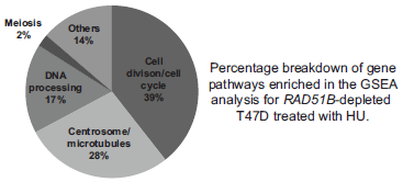
RAD51B depletion leads to changes in gene expression post DNA damage. Gene set enrichment analysis (GSEA) results for three independent experiments are presented. DNA microarray analyses were performed to compare gene expression in T47D cells transfected with siRNA targeting RAD51B or non-targeting siRNA control, both observed 24 hours after 2 mM HU treatment. Graph depicts percent breakdown of pathways which contain genes enriched within RAD51B-depleted populations.
To validate the gene expression results, commercial qPCR-based expression arrays were used to detect expression changes in 80 DNA-damage response genes. Ten genes showed significantly altered expression, with fold changes averaging between 2 and 14 (p < 0.05). Of the 10 genes, four are involved in cell cycle checkpoint-related processes, and the rest are involved in DNA repair pathways (Table 1).
qPCR-array based gene enrichment analysis was performed to verify GSEA results. Table shows 10 genes with significant changes (p value <0.05) between T47D cell populations depleted of RAD51B and control post HU treatment.

Depletion of RAD51B leads to differential cell cycle arrest in response to DNA damage agents
In order to determine whether RAD51B protein levels alter the cell cycle checkpoint response to DNA damage, we investigated expression patterns for RAD51B protein during the cell cycle after cell synchronization. We did not observe fluctuation of protein levels across cell cycle phases (Fig. 5A). To provide evidence that RAD51B depletion altered cell cycle checkpoint regulation in response to DNA damage in breast cancer cells, we analyzed the cell cycle progression profile following exogenous DNA damage induced by CDDP and HU using the same conditions as the drug sensitivity experiments (Fig. 5B). T47D cells transfected with siCON or siRAD51B exposed to 1 mM HU for 72 hours or 100 uM CDDP for 24 hours were analyzed as were BT549 cell transfected with siCON or siRAD51B treated 72 hours with 1 mM HU (Fig. 5B). We observed differences in the response to CDDP and HU exposure between RAD51B-depleted and control cell samples. We observed 3.07% more cells arrested in the G2/M phase in the RAD51B-depleted samples compared with controls in the CDDP exposure experiment (p = 0.03), and 15.4% fewer cells in the S phase of the cell cycle for the RAD51B-depleted samples in the HU exposure experiment (p = 0.03). A similar cell cycle arrest profile was observed in RAD51B-depleted BT549 cells post HU treatment, in which 8.0% fewer cells were observed in S phase compared with controls (p = 0.01) (Fig. 5C). These findings suggest that RAD51B depletion disrupts the normal cell cycle response to CDDP and HU-induced DNA damage.

Effects of DNA damage on cell cycle progression in RAD51B-depleted breast cancer cells. (A) RAD51B protein expression during the cell cycle in T47D breast cancer cells. T47D cells were synchronized at G0/G1 phase by serum starvation for 48 hours. The cells were harvested at indicated time points post serum stimulation and analyzed to determine the DNA content by propidium iodide staining and FACS. Graph represents portion of cells in G1, S, or G2/M at each time point. Experiments were repeated three times. Western blot analysis was performed to detect RAD51B protein levels at each time point. (B) Representative figure of cell cycle distribution of T47D and BT549 cells with (siRAD51B) or without (siCON) RAD51B depletion and treated with 1 mMHU for 72 hours. (C) Graph reports average cell cycle distribution of three independent experiments.
Discussion
The function of RAD51B in breast cancer cell lines has not been well studied and most previous work has been reported in non-mammary cells. Herein, we examined the response to DNA damage in breast cancer cell lines in which RAD51B expression was reduced by siRNA and observed an increased sensitivity to DNA-damaging agents in two of three cell lines studied; specifically, enhanced sensitivity to CDDP and HU in T47D cells, and increased HU sensitivity in BT549 cells (Figs. 1B and C). T47D cells are ER-positive, PR-positive, and HER2-negative and are responsive to chemotherapy. On the other hand, BT549 cells are triple negative and show only an intermediate response to chemotherapy. Since these cell lines represent distinct subtypes of breast cancer, 44 it is plausible that a reduction in RAD51B could contribute to carcinogenesis differentially in distinct subtypes of breast cancer. Overall, our data suggest a reduction of RAD51B in breast cancer cell lines leads to increased sensitivity to DNA-damaging agents, similar to what is observed in non-mammary cells. 35 Interestingly, we did not observe comparable results with the WST-1 viability assay, which is predicated on metabolic activity only, and the colony formation assay, which requires cell survival and proliferation. It has been reported that compared with the clonogenic assays, cell proliferation-based assays yield smaller differences in the surviving fraction between the control and DNA-repair defective breast cells after identical radiation treatment, 45 which could partially explain the inconsistency between the assays.
Next, we investigated the possible effect of RAD51B on I-SceI generated DSB induced HR in the modified breast cancer cell line MCF 7 (Fig. 2) and observed comparable results to a previous report. 46 In this regards, reduction of HR efficiency may partially explain the increased DNA damage sensitivity. Thus, one of the functions of RAD51B in breast cancer cell lines could be to protect the genome against DNA damage through its participation in the HR repair pathway.
In response to DNA damage, we measured RAD51B mRNA and protein levels in breast cancer cell lines treated with CDDP, HU, bleomycin, and MMS, since each induces DNA repair by different mechanisms. The bulk of DNA adducts formed in response to CDDP exposure are intrastrand cross-links repaired by the trans-lesion synthesis polymerases, allowing error-prone bypass of the lesion. The inter-strand cross-links, which constitute a smaller portion of CDDP damage, are repaired by several mechanisms including the Fanconi's Anemia and HR pathways. 47 Prolonged treatment with HU can stall the replication fork 48 and the HR pathway is one method for restarting the replication process. 49 Bleomycin induces single strand breaks and DSBs as the result of base-elimination from the nucleic acid strands, 50 whereas MMS modifies DNA by adding methyl groups to bases repaired initially by the base excision repair (BER) pathway. DNA-adducts that are not repaired lead to stalled replication forks that are subsequently repaired by HR.51,52 We observed that RAD51B transcript levels decreased in T47D and MCF7 cells treated with MMS or HU (Fig. 3A). Interestingly, we observed elevated protein expression in MCF7 and BT549 cells at the 5 hour recovery time post HU-treatment (Fig. 3B). Although this increase in RAD51B protein level is in line with a previous report, 33 we failed to observe similar changes at the transcriptional level. These data suggest that post DNA damage, RAD51B levels may be largely regulated at the level of translation or post-translation. The differences observed between the three cell lines are consistent with prior observations for other DNA repair and replication-associated genes. For example, RAD51 gene expression is altered by different transcriptional activators and repressors,53,54 but it is not induced uniformly by DNA damage agents, such as CDDP, MMS, and UV treatment.55,56 Similarly, DNA damage is reported to cause no changes or only a slight decrease in RAD51 mRNA or protein levels in different cell lines.57–59
The increased sensitivity to DNA-damaging agents in RAD51B-depleted cell lines could be due to disruption of cell cycle progression. To investigate this possibility, FACS analysis (Figs. 5B, C) showed that depletion of RAD51B prior to treatment with CDDP resulted in an increased arrest of cells at G2/M. This is consistent with data from CHO hamster and L11210/0 mouse leukemia cells, which showed that CDDP-induced lethality results primarily in G2 cell cycle arrest and subsequent apoptosis; furthermore, they also reported cells deficient in NER experienced a longer G2 arrest than repair-proficient cells.60,61 A recent study in yeast showed that trans-lesion synthesis and HR are redundant during G2 for the repair of single strand DNA gaps. 62 Exposure to HU induced a higher fraction of cells arrested in S phase in normal T47D cells, but not in RAD51B-depleted cells consistent with the observation that overexpression of the phosphatase CDC25A led to depression of G1/S cell cycle arrest in response to HU treatment, elevation in premature mitosis, and subsequent cell death. 63 It is possible that the lack of S phase delay upon HU treatment in RAD51B-depleted cells prevented proper repair of stalled replication forks, resulting in increased cell death similar to the observation in cells overexpressing CDC25A.
Gene expression patterns were investigated in breast cancer cell lines depleted of RAD51B by siRNA and subjected to prolonged treatment with HU. Commercially available DNA repair pathway qPCR arrays were used to confirm expression patterns, with high concordance. Our results indicate that depletion of RAD51B reduced expression of genes involved in cell division/cell cycle by GSEA analysis. Notably, we observed altered expression of CHEK1, CHEK2, RAD1 as well as replication genes, DDB2, FEN1, in addition to those involved in centrosome/microtubule processes. We also observed disruption of expression of genes critical for repair pathways, namely, NER (DDB2), BER (UNO), HR (MRE11, RBBP8), and Fanconi Anemia (FANCD2, FANCG) (Fig. 4; Table 1). These data suggest that RAD51B depletion alters transcription of genes involved in DNA repair and cell cycle checkpoint response pathways, consistent with effects seen in the treated cell lines.
There are at least two alternative explanations, either (1) HU treatment generated stalled replication forks during S phase of the cell cycle due to reduced HR-repair subsequent to RAD51B-knockdown as HU causes stalled replication forks by depleting the pool of certain nucleotides that are used to extend the replicating strands, or (2) RAD51B may directly influence the cell cycle by interacting with a component of the checkpoint response pathway, similar to the interaction observed between BRCA1 and cell cycle regulation proteins. 64 Based on the expression data under conditions that did not lead to significant differences in viability/proliferation, as well as a previous study on RAD51B protein foci formation in response to platinum drug treatment, 39 we believe it is highly likely for RAD51B to interfere with the cell cycle checkpoint independent of its role in HR repair. However, further work is needed to confirm this interpretation.
Conclusion
We report here on studies of the biological function of RAD51B in breast cancer cell lines as a first step in the investigation of the mechanism underlying genetic variants of RAD51B that are associated with breast cancer risk. RAD51B depletion enhances sensitivity to DNA damage agents, reduces HR repair function, and is involved in cell cycle checkpoint response. In light of the complex interaction between repair and cell cycle checkpoint responses, it will be critical to investigate the mechanism by which RAD51B influences cell cycle checkpoint response, particularly in breast cancer cells in relation to the common genetic haplotypes that confer susceptibility to breast cancer.
Author Contributions
Responsible for concept, design, analysis, and interpretation of experiments as well as preparation of a draft manuscript: PSL. Performed FACS experiments: JF. Performed western blots, assisted in interpretation of experiments, and performed critical revisions of the manuscript: LJ. Performed western blots, and assisted in revising the manuscript: TM. Drafted critical revisions of the manuscript: PR. Performed the gene expression microarray experiments: NH, CW, and PRT. Performed the GSEA pathway analysis: JW and JK. Provided MCF7-DFP cells and pCBASce plasmid, reagents essential for this work: MJ. Responsible for the concept and interpretation of experiments as well as critical revisions of the manuscript: SJC. All authors read and approved the final manuscript.