
Abstract
A sensitive and reliable method of liquid chromatography–electrospray ionization/tandem mass spectrometry (LC-ESI/MS/MS) was developed and validated for determining 1,3-dimethylamylamine (1,3-DMAA) and 1,4-dimethylamylamine (1,4-DMAA) in geranium plants (
Keywords
Introduction
Dimethylamylamine (DMAA), also known as methylhexaneamine, or 1,3-dimethylpentylamine, is a simple aliphatic amine (Fig. 1). In recent years, it has appeared as an ingredient in various dietary supplements and sports nutrition products. This use of the compound has given rise to inquiries as to whether DMAA is a naturally occurring constituent of the geranium ( Structures of 1,3-Dimethylamylamine (1) and 1,4-Dimethylamylamine (2).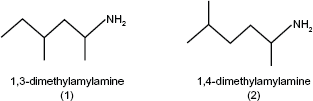
The result of a GC/MS analysis of geranium oil, which indicated the presence of 1,3-DMAA (1) and 1,4-DMAA (2) was published in a journal that has not been widely circulated internationally. 1 Aside from that publication, geranium essential oil has been the subject of numerous other investigations seeking to identify and quantify all of the compounds present, generally employing the use of GC/FID and GC/MS; these other investigations have failed to identify 1,3-DMAA (1) or 1,4-DMAA (2).2–12 However, because geranium oil presents such a complex sample matrix and contains many volatile compounds, a possible shortcoming of the GC/FID method is interference by the sample matrix. 6 Although the use of GC/MS is an improvement, the complex nature of the sample matrix still presents issues when seeking to identify all components of the oil; 6 in fact, we are not aware of any published data demonstrating an identification of components comprising 100% of the oil. These factors, along with differences in sample origin, processing and composition, may explain why these other investigations failed to identify DMAA as a component of geranium oil. Regarding 1,3-DMAA (1), it has been noted that although it is amenable to GC/MS analysis, 13 great care must be taken during method development because of DMAA's strong polarity, volatility and low molecular mass, making it a challenge for GC-column retention and separation.
DMAA has been successfully determined in urine using LC/MS/MS.13,14 In those investigations, 1,3-DMAA (1) was detected as a pair of peaks with identical MS/MS spectra. However, the determination of DMAA in plant and especially in geranium oil has yet to be published. In the present study, we have developed and validated a sensitive and simple LC/MS/MS method to identify 1,3-DMAA (1) and 1,4-DMAA (2) and determine their concentrations as naturally occurring compounds in geranium plants.
Experimental
Chemicals and reagents
1,3-Dimethylamylamine (1) (CAS No. 105-41-9, >99%) and 1,4-dimethylamylamine (2) (CAS No. 28292-43-5, >99%) were purchased from Sigma-Aldrich (St. Louis, MO, USA). Formic acid (88%), methanol (HPLC grade), hexane (pesticide residue grade) and hydrochloric acid (37% HCl) were all obtained from Fisher Scientific (Waltham, MA, USA). All other chemicals are analytical grades. Water (18.2 MΩ-cm) was prepared with a Barnstead NanoPure Diamond System (Lake Balboa, CA, USA).
Apparatus and instruments
Agilent 1100 HPLC system with quaternary pump (Santa Clara, CA, USA) was coupled to a Micromass Quattro Ultima mass spectrometer with electrospray ionization (ESI) source (Manchester, England). Masslynx (version 4.1) were used to control the system of LC-triple quadrupole mass spectrometer and for data acquisition and processing. The analytical column is a Phenomenex Kinetex C18 column (4.6 * 150 mm, 2.6 μm) (Torrance, CA, USA). High Speed Grinder DFY-200 was from Gaoyi In. (Wenzhou, China). Commercial Blender 200G was from Waring Co. (Torrington, CT, USA).
Geranium plants and geranium oils
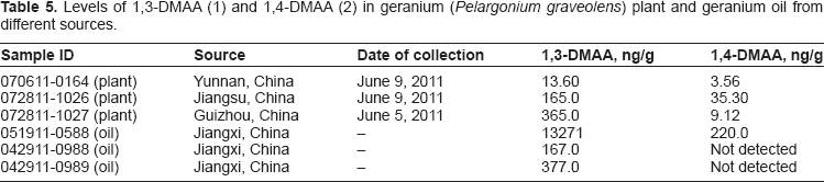
The geranium plants were procured by and obtained from Dr. Yi Jin of Yunnan University (Kunming, Yunnan Province, China) and were authenticated by Professor Xu Youkai of the Xishuangbanna Tropical Botanical Garden-Chinese Academy of Sciences (Mengla, Yunnan Province, China). The plant samples were collected from different areas of China (
Levels of 1,3-DMAA (1) and 1,4-DMAA (2) in geranium (
Standard preparation
The stock solutions of 1,3-DMAA (1) and 1,4-DMAA (2) were prepared separately in methanol at a concentration of 1.00 mg/mL and 1.068 mg/mL, respectively. Working solutions composed of the two DMAAs were prepared by serial dilution of the stock solution with 0.5 M HCl to obtain a set of standard concentrations of 0.10-10.00 ng/mL of 1,3-DMAA (1), and 0.11-10.68 ng/mL of 1,4-DMAA (2). All working standard solutions were stored at 4 °C and used within one week after preparation, although no significant degradation was observed in one month of storage.
Sample preparation and extraction
Geranium plant (wet leaves and stems) was thawed at room temperature and cut into pieces at about 1-2 cm and mixed well. After about five hundred grams of the plant was ground into fine pieces with a high speed grinder, 10 g of sample was weighed into a stainless steel blender cup. To this cup, 80 mL of 0.5 M HCl was added and mixed. The mixture was homogenized at high speed for 2 min. The homogenate was then transferred into a 100-mL volumetric flask. The blade and cup were washed with additional 15 mL of 0.5 M HCl. The solution was collected into the flask and extracted by sonication at 50 °C for 1 hour. After being cooled to room temperature, the volume was adjusted to the mark with 0.5 M HCl. This solution was centrifuged at 4000 × g for 10 min, and the supernatant was further purified as below.
For geranium oil, 1 mL of sample was mixed with 1 mL of hexane in a 10-mL glass tube with screw cap. Five mL of 0.5 M HCl was added and shaken with a vortex shaker for 5 min at high speed. The aqueous layer (lower) was diluted with 0.5 M HCl as necessary, filtered with a 0.45-μm nylon filter and applied to LC/MS/MS without further purification.
Purification
Four mL of supernatant and 2 mL of hexane were added to a 10-mL glass tube with screw cap. The mixture was shaken by a vortex shaker for 30 sec. The mixture was centrifuged at 2000 × g for 5 min. The aqueous layer was diluted as necessary and filtered for LC/MS/MS analysis.
LC/MS/MS conditions
The mobile phase of HPLC is composed of Water:acetonitrile (85:15) containing 0.1% formic acid. Flow rate was 0.5 mL/min; column temperature was 35 °C; the flow was diverted 0.2 mL/min to MS. Injection volume was 50 μL.
The mass spectrometer was operated in positive ESI and multiple reaction monitoring (MRM) mode. Nitrogen was used as the nebulizer, heater, and cone gas. Argon was used as the collision induced dissociation (CID) gas. The precursor-to-product ion transitions were monitored at m/z 116 [M + H] → 57 (quantification) and m/z 116→99 (qualification) for both 1,3-DMAA (1) and 1,4-DMAA (2). ESI parameters were optimized for maximizing the generation and stability of the precursor and fragment ions by infusion as follows: Capillary 2.5 kV, Cone 20V, Source temperature 120 °C, Desolvation temperature 360 °C, Cone gas 120 L/hour, Desolvation gas 850 L/hour, CID 11 eV, collision cell pressure 2 × e-3 mbar.
Method validation
The analytical method was validated according to guidelines for United States Pharmacopeia (USP). The parameters validated include linearity, specificity, limit of detection, limit of quantification, accuracy, precision and reproducibility.
Linearity
To evaluate the linearity, calibration curves of 1,3-DMAA (1) and 1,4-DMAA (2) were established using concentrations in the range from 0.1 to 10 ng/mL. The responses of each compound against its respective concentration were plotted. Linear regression analysis was performed to obtain calibration equation and correlation coefficients (R 2 ).
Specificity
Two ion transitions coupled with a high resolution column were used to enhance the method selectivity. Specificity was assessed by comparing the chromatograms of blanks (glassware and reagent blanks), standard, spiked samples and their peak purity (peak shape and relative intensity of transitions). The peak was identified by retention time and relative intensity of transitions against the reference standard.
Matrix effects
The matrix effects (ion suppression or enhancement) were evaluated by comparing peak area of the standard, sample extract and the extract directly spiked at corresponding concentrations of DMAAs, which were set at the medium spiked concentration (20 ng/g).
Accuracy
Accuracy of the method was determined by assaying spiked samples of geranium plants at four different levels: about 5, 10, 20 and 40 ng/g for both 1,3-DMAA (1) and 1,4-DMAA (2) (ie, adding 50, 100, 200 or 400 ng DMAA in solution to about 10 g of sample). Each concentration level had three replicates. All samples were extracted, purified and determined as described above. For the blank, 10 mL of water instead of sample was included and carried through the same procedures of sample preparation.
Precision and reproducibility
Precision was performed by assaying a geranium sample in six subsamples. The concentration of each subsample, and average and RSD of the analyses were calculated to access the precision of the method.
Reproducibility of this method was evaluated by a second chemist beside the primary chemist in this laboratory. The geranium sample was assayed in six subsamples. The concentrations, average and RSD of the six analyses were compared with the results obtained by the primary chemist to assess the reproducibility of the method by a different chemist.
Data analysis
The concentrations of 1,3-DMAA (1) and 1,4-DMAA (2) in the sample preparations were obtained from their corresponding standard curves. The mean, standard deviation (SD) and relative standard deviation (RSD) of spike recoveries were calculated for assessing the accuracy of the method. Mean and RSD of repeated analyses of the geranium samples were used for precision evaluation. The mean concentration of DMAAs from the precision experiments was used as the original value to calculate recoveries of DMAAs. The recoveries from spiked samples were calculated by the following formula:

where, Fc is the concentration (ng/g) found in the spiked sample; Bc is the original value of the sample (ng/g) prior to spiking; Sc (ng/g) is the concentration spiked to the sample.
Results and Discussion
Analytical conditions
Because DMAA is a simple aliphatic amine with no chromatic group, the HPLC-UV method is not suitable for its detection unless the compound is derivatized with a chromophore prior to analysis. Therefore, LC/MS/MS was selected for the current method. Two solvent systems, methanol-water and acetonitrile-water were tested for optimization on the C18 column and tuning of ESI/MS. Both solvent systems produce similar ESI (+) signals. Further investigations showed that formic acid (0.1%) in either mobile phase enhances the signal significantly, with little effect on retention time and shape of the peaks. As a result, acetonitrile:water (15:85) containing 0.1% formic acid was selected and used routinely as mobile phase in the current experiment. Under these conditions, 1,3-DMAA (1) and 1,4-DMAA (2) were well separated with good peak shape (tailing factor = 1.1-1.3) and retention (K’ = 2.5-2.8) (Fig. 2).
MRM chromatograms of (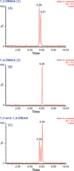
When a standard solution of 1,3-DMAA (1) was analyzed under the current conditions, double peaks with similar intensity were observed. Similar results were observed by Vorce et al
13
and Perenoud et al.
14
Both peaks show identical CID mass spectra (data not shown), suggesting that these peaks are stereo isomers (Fig. 2A). In contrast, 1,4-DMAA (2) shows only a single peak (Fig. 2B). 1,3-DMAA (1) has two chiral centers (carbon-1 and carbon-3) in its structure, and thus it theoretically has four stereo isomers. The double peaks observed are likely formed by its diastereomers, (1
Figure 3 shows that 1,3-DMAA (1) and 1,4-DMAA (2) produced similar CID mass spectra for the same precursor ion m/z 116 [M+H]+). Their CID spectra had a strong and stable product ion m/z 57 [C4H9]+ that was used for quantification. Other product ions with relatively higher abundance were m/z 43 [C3H7]+, m/z 75 and m/z 99 [M+H-NH3]+.
The CID spectra (11 eV) of 1,3-DMAA (1) (Top); 1,4-DMAA (2) (Bottom).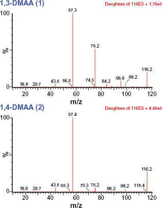
Sample preparation, matrix effect and specificity
DMAA is slightly soluble in water, but soluble in diluted HCl and many polar solvents. Both 0.5 M HCl and methanol were examined for their extraction of the geranium samples. It was found that both solvents had similar extraction efficiency. However, methanol extracts much more of the matrix components from the geranium plant than 0.5 N HCl. The final extract prepared using methanol was found to interfere with LC/MS analysis (data not shown). In contrast, the extract prepared using 0.5 N HCl carries less matrix components, especially fat-soluble compounds, which was later found to be important for the accuracy of determination. Therefore, the diluted HCl solution was used for routine sample extraction. Furthermore, sonication for 60 minutes of the geranium sample was found to be necessary for reproducible results.
The recovery of DMAAs in initial experiments was found to be low (about 60%-70%) when the HCl-extract was directly applied to LC/MS/MS without further purification. To investigate the cause of this low recovery, our focus was on the effect of the sample matrix, since the ESI signal is more susceptible to matrix-induced suppression. Results of the investigation clearly showed that the matrix of the sample had suppressed the signal of DMAAs in the sample extract or spiked sample extract (Fig. 4). For example, after subtracting the background, the ESI signal of 1,3-DMAA (1) in the spiked extract with purification was about 35% greater than that without purification (Fig. 4B and C). These results suggested that ion suppression had occurred in the raw extract. It is also noted that ion-suppression is slightly less at the elution window for 1,4-DMAA (2), which was only 20% lower in the spiked extract without purification.
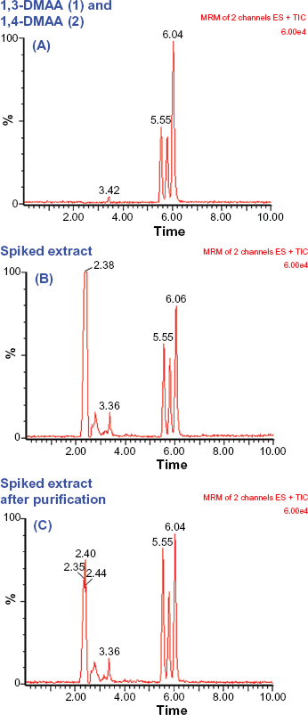
Matrix effect on determination of DMAAs in geranium (
It has been known that ion suppression occurs in many ESI/MS-based methods for biological samples. 16 Methanol is a strong protic solvent and can solubilize considerable amounts of lipid components, such as fatty acids and phospholipids. Considering the ESI mechanism, these nonvolatile or less volatile compounds were the potential solutes suppressing the ESI signal. 17 The diluted HCl solution used in the current method provides strong solubility for DMAAs but solubilizes considerably less of these fat-soluble components. Furthermore, the HCl-extract is readily purified by partitioning with hexane to remove the lipid-soluble components. This simple purification step was found to be effective in reducing sample matrix and ion suppression, and improved recovery significantly (Fig. 4B and C).
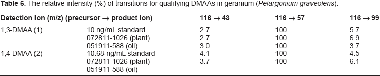
The relative intensity (%) of transitions for qualifying DMAAs in geranium (
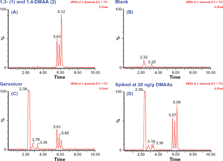
MRM chromatogram of (
Linearity
Standard solutions of 1,3-DMAA (1) and 1,4-DMAA (2) were analyzed with concentration range at about 0.1 to 10 ng/mL. The two isomer peaks of 1,3-DMAA (1) was summed prior to regression analysis. The results were linear with R 2 of 0.998 for 1,3-DMAA (1) (Y = 2537X + 497.6, Y-peak area, X-ng/mL), and 0.999 for 1,4-DMAA (2) DMAA (Y = 2705X + 311.3).
Precision
Precision for determination of 1,3-DMAA (1) and 1,4-DMAA (2) in geranium (
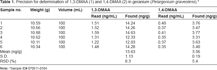
Note:
Sample ID# 070611-0164.
Accuracy
Accuracy for determination of 1,3-DMAA (1) in geranium (
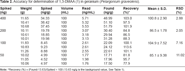
Note:
Recovery (%) = (Found-13.63)/Spiked × 100 (13.63 ng/g is the background value, See Table 1).
Accuracy for determination of 1,4-DMAA (2) in geranium (
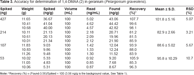
Note:
Recovery (%) = (Found-3.56)/Spiked × 100 (3.56 ng/g is the background value, See Table 1).
Detection limit and quantification limit
The instrument detection limit was estimated by analyzing a standard at a concentration of 0.2 ng/mL with injection volume of 50 μL. The chromatogram is shown in Figure 6. The detection limit was estimated to be 1-2 pg, based on the signal-to-noise ratio of 3:1.
MRM chromatogram for estimating the instrument detection limit: 0.20 ng/mL 1,3-DMAA (1) (5.5 min and 5.8 min) and 0.11 ng/mL 1,4-DMAA (2) (6.0 min).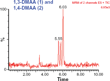
To evaluate the method quantification limit (MQL), the signal to noise ratio of 5:1 is used for calculation. Taking into consideration of the sample weight of 10 g and the final volume of sample preparation in 100 mL, the MQL of 1,3-DMAA (1) and 1.4-DMAA (2) is estimated to be 1-2 ng/g.
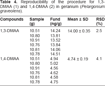
Reproducibility
Reproducibility of the procedure for 1,3-DMAA (1) and 1,4-DMAA (2) in geranium (
Application of the method to investigating geranium plants and geranium oils
The current method was applied to analyze geranium plants and geranium oils from different sources. The results are shown in Table 5. As expected, 1,3-DMAA (1) was further confirmed by multi-ion transition and the product ion ratios (Table 6). These results provide strong evidence that 1,3-DMAA (1) and 1,4-DMAA (2) are naturally present in both geranium plant and geranium oil.
A major advantage of the currently described method enabling detection of DMAAs in geranium plants and oils is its extreme sensitivity. The instrumental detection limit is approximate 1 pg. Another advantage is its simplicity. The method involves simple sample extraction and sample partition with hexane. There are no extended purification and derivatization steps involved, which should be necessary to GC/MS. The accuracy and precision of the method at ppb levels is easily achievable with a conventional LC column and mobile phase.
While the presence of DMAA has been reported in geranium in one investigation, those data were not considered conclusive due to issues regarding the experimental design and data analysis. 1 Therefore, to our knowledge, the present study is the first to show conclusively that DMAA is naturally occurring in geranium plants.
It is well-documented that the variations of environmental conditions and geographical locations have great effect on the chemical profiles of the geranium plant.5,8 The results in the current study showing various amounts of DMAA in geranium plants from different regions appear to be consistent with these observations (Table 5). Although the proportion of 1,3-DMAA (1) to 1,4-DMAA (2) varied considerably from sample to sample, in general, the concentration of 1,3-DMAA is much higher than that of 1,4-DMAA (2), suggesting that 1,3-DMAA (1) is the predominant form naturally occurring in geranium plants. The fact that 1,3-DMAA (1) is highly concentrated in one geranium oil sample when compared to the other two geranium oils could have been a result of either different geranium plants used for oil processing or from different manufacturing processes.
Another intriguing observation is that 1,4-DMAA (2) was not detected (below quantification limits) in two geranium oil samples where 1,3-DMAA (1) was present although at relatively lower concentrations. This discrepancy was unexpected. One explanation might be that 1,4-DMAA (2) is not stable at low concentrations. However, based on the structure of 1,3-DMAA (1) and 1,4-DMAA (2), they appear to be stable molecules with relaxed structure and no labile parts under various storage temperature conditions. Thus, an alternative explanation is that these two samples contained a higher ratio of 1,3-DMAA (1) to 1,4-DMAA (2). We have noted in the other samples, varying 1,3-DMAA (1):1,4-DMAA (2) ratios of approximately 5:1, 40:1 and 60:1, thus it is possible that with a combination of an even higher ratio and a smaller amount of 1,3-DMAA (1) present, the 1,4-DMAA (2) would have been below our quantification limits.
The results from the present study show that 1,3-DMAA (1) has two isomer peaks which are present in equal amounts and which are identical in all tested samples, including the standard reference. The reference standard of 1,3-DMAA (1) is synthetic and produced via chemical reaction. However, most compounds present in plants should be made through an enzymatic process. Therefore, most likely only one chiral configuration would be present in plants (often referred to as natural form). The results in the current study show that 1,3-DMAA (1) in geranium plants and geranium oils appears to be an exception to this notion. Indeed, this is not the first report demonstrating the presence of a racemate in a plant tissue.18–20 In fact, the presence of a racemate (ie, nerol oxide) has been demonstrated once before in the geranium plant as well. 19 Further study is needed to elucidate the biosynthetic pathway of DMAAs in the geranium plant.
Conclusion
DMAA, which is used in some nutritional supplements, has led some to question whether it is actually a constituent of the geranium plant and its oils. A validated method for quantification of DMAA in geranium plants has been established in the present investigation and has confirmed the presence of 1,3-DMAA (1) and 1,4-DMAA (2) in the plant tissue and essential oil. The conditions of LC and ESI positive MS/MS have been optimized. A simple and rapid procedure for sample extraction and purification has been developed. This LC/MS/MS method is sensitive and reliable and has been used successfully for the simple and rapid analysis of DMAA in the geranium plant and its oils.
Author Contributions
J.S. Li and M Chen were responsible for data collection/analysis; J.S. Li was primarily responsible for manuscript preparation. Z.C. Li was responsible for study design and revision of manuscripts. All authors have read and approved the final manuscript.
Funding
This research was financially supported by USPlabs LLC.
Competing Interests
JSL, MC and ZCL disclose that funding for analytical research and manuscript preparation was provided by USPlabs, LLC. ZCL served as an expert witness in 2011 for the case: DeRosier v. USPlabs. Assistance with English grammar in preparation of the manuscript was provided as a courtesy to the authors by The Brewer Law Group, PLLC.
The sponsor initiated a request to this laboratory to investigate the presence of DMAA in geranium plant and geranium oil. All experimental design, method of extraction and method of quantification were carried out independently by Intertek-AAC Labs. Data analysis and the manuscript preparation were performed by the authors of the manuscript, while the sponsor provided grammatical review and assistance. The submission of the paper for publication was suggested by Intertek-AAC Labs to the study sponsor and the sponsor agreed.
Footnotes
As a requirement of publication author(s) have provided to the publisher signed confirmation of compliance with legal and ethical obligations including but not limited to the following: authorship and contributorship, conflicts of interest, privacy and confidentiality and (where applicable) protection of human and animal research subjects. The authors have read and confirmed their agreement with the ICMJE authorship and conflict of interest criteria. The authors have also confirmed that this article is unique and not under consideration or published in any other publication, and that they have permission from rights holders to reproduce any copyrighted material. Any disclosures are made in this section. The external blind peer reviewers report no conflicts of interest.